

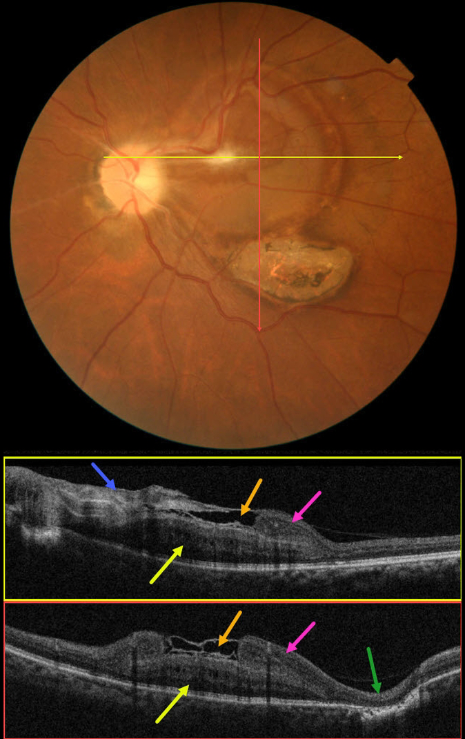
This 61YO male presented with 16 days of blurred vision and floaters in his left eye. There was no prior ocular history, and the past medical history was normal. Vision was 20/30 in his normal right eye and 20/200 in his left eye.
Color imaging shows an unusual circular area of subretinal yellow-orange pigmentary changes extending temporally from the optic nerve throughout the central and superior macula. A contracted localized area of premacular fibrosis is noted in the superonasal fovea. A flat area of variably pigmented chorioretinal scarring is found inferotemporally.
OCT B-scans show a constellation of interesting findings. The nasal epimacular fibrosis extends temporally, giving the appearance of either an ILM drape or local separation over the macular center. More temporally disorganized hyperreflective tissue descends and seems to merge into definite retinal inner tissue. There is complete outer retinal loss overlying the inferotemporal area of scarring.
The deeper retina is occupied by a mound-like area of alternating hyper- and hyporeflective bands. It is unclear whether this represents thickened outer retina or an unusual scar, as it appears distinct and anterior to the well-visualized ellipsoid zone below.
We believe this constellation of findings most likely represents changes following toxoplasmosis chorioretinitis, but we also can’t rule out an independent combined hamartoma. Or maybe we’re dealing with another diagnosis. What do you think?



