

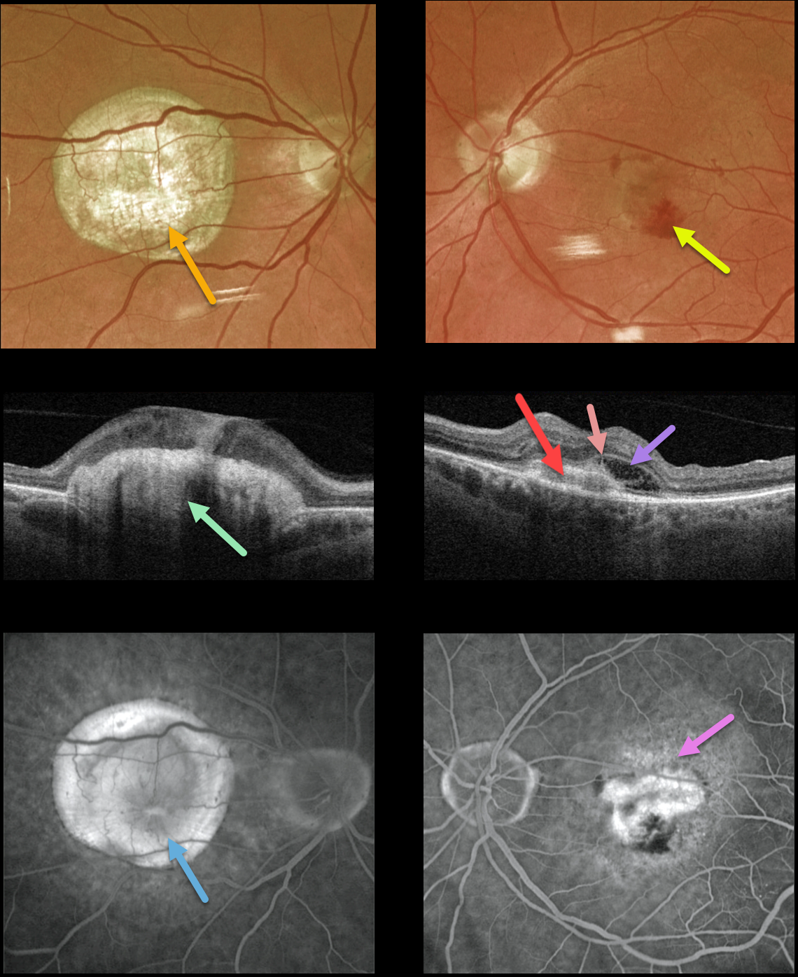
This 77YO male presented with counting fingers vision OD from a large fibrotic disciform scar and 20/200 vision OS from a new macular neovascularization.
The disciform scar is contracted and fibrotic, with marked hyperreflectivity on swept-source OCT. The MNV in his left eye is seen as opaque fluid with some subretinal blood. The type 2 MNV (located above the RPE) is noted on the OCT B-scan. There is also a bacillary detachment and a hyperreflective line indicating a possible pitchfork sign.
Optos fluorescein angiography shows diffuse staining of the subretinal scarring OD and leakage from the MNV OS.
Learning Points:
Disciform scars were the norm for untreated wet AMD before the anti-VEGF era that began in the mid-2000s. New-onset wet AMD diagnosed and treated with appropriate anti-VEGF dosing virtually never goes on to develop these lesions. There may be progressive macular atrophy following years of therapy, but not this type of fibrotic scarring.
An excellent review of wet AMD fibrotic lesions is in Souied et al’s AOS thesis (AJO 2020;214:151-171).
The pitchfork sign was originally described by Hoang et al (Retina 2013;33:1049-1055) as a unique OCT finding in inflammatory MNV. Falavarajani et al more recently noted that this finding is also associated with non-inflammatory MNV (Ophthalmic Surg Lasers Imaging Retina 2019;50:719-725).
The pathogenesis of the avascular pitchfork spikes is unknown, but may be related to outer retinal traction or Müller cell activation.



