

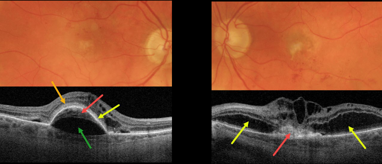
This 87YO patient presented with vision of 20/200 OD and counting finger vision OS from bilateral active neovascular AMD (nAMD).
The right macula has a few dots of central and superior macular blood. OCT scanning shows a type 1 macular neovascularization (MNV, located below the RPE) adherent to an overlying RPE detachment. A prechoroidal cleft separates the MNV from the underlying choroid. Subretinal hyperreflective material (SRHM) lies above the RPE detachment (orange arrow). Intraretinal fluid is noted nasally with trace subretinal fluid on either side of the RPE detachment.
The left macula has faint subfoveal fibrosis with some scattered more inferior subretinal blood. OCT scanning shows a type 2 MNV (located above the RPE), which is tethered to the overlying edematous retina. Bacillary layer detachments are noted nasal and temporal to the MNV.
Learning Points:
A prechoroidal cleft is a hyporeflective space between the RPE and Bruch’s membrane. These are seen in up to about 20% of treated eyes with nAMD, particularly with type 3 neovascularization (intraretinal MNV) and polypoidal choroidal vasculopathy.
They are usually associated with worse visual acuity due to potential complications, including RPE tear and subretinal hemorrhages (Kim et al, Retina 2017;37:2047-2055).
SHRM is an OCT finding that is invisible clinically. This material, located between the neurosensory retina and retinal pigment epithelium (RPE), is thought to consist of fluid, fibrin, blood, and other fibrovascular tissues.
SHRM is a biomarker of active disease that decreases with successful anti-VEGF therapy (Willoughby et al., Ophthalmology 2015;122:1846-1853). Persistent SHRM is associated with an increased incidence of scar formation and worse VA.
Bacillary layer (cone and rod inner and outer segments) detachment appears as a unique dome-shaped collection of intraretinal fluid from photoreceptor splitting. These detachments can be observed in an increasing number of conditions, most classically Vogt-Koyanagi-Harada disease (Cicinelli et al, Ophthalmology Retina 2020;4:454-456).



