

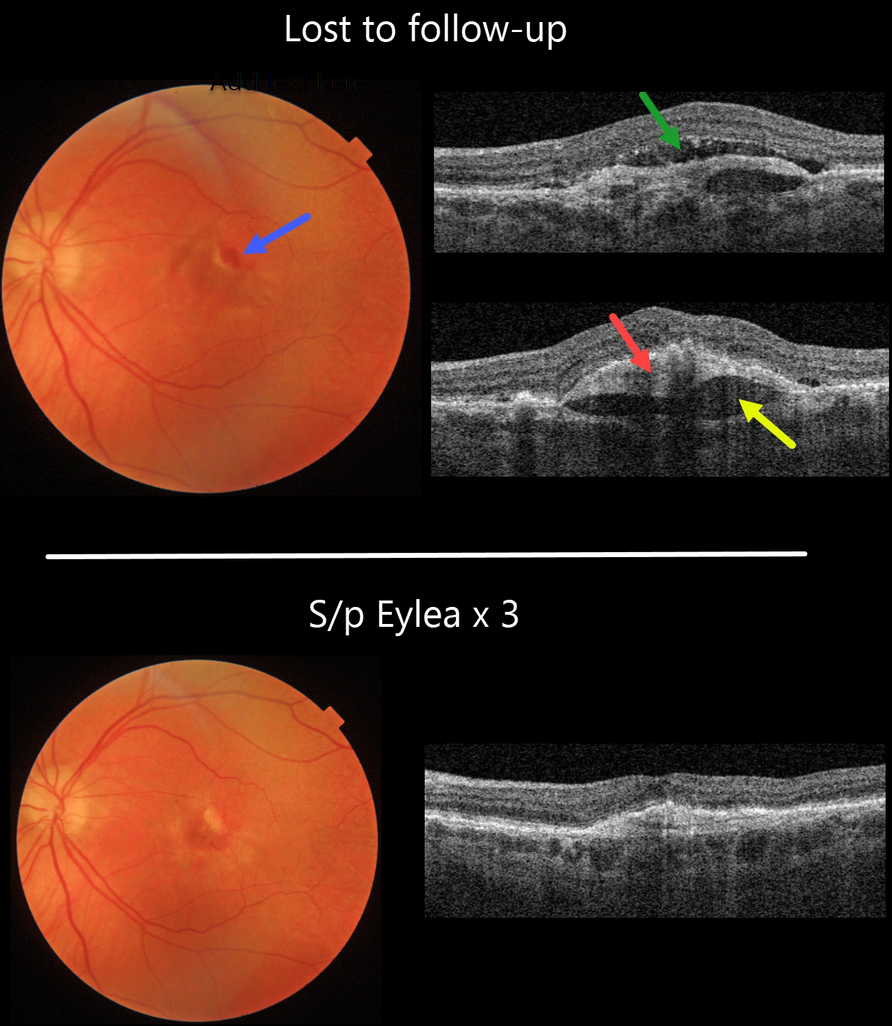
This 82YO female was maintaining 20/40 vision in her left eye with intravitreal Eylea every 8 weeks for neovascular AMD. She was then lost to follow-up, with new blood and 20/50 vision.
Swept source OCT shows new subretinal fluid. The hyperreflective sub-RPE type 1 macular neovascularization is elevated above a hyporeflective prechoroidal cleft.
Fortunately, vision returned to 20/30, with complete resolution of all exudative findings, following 3-monthly Eylea injections.
Learning Points:
A prechoroidal cleft is a hyporeflective space between the RPE and Bruch’s membrane. These are seen in up to about 20% of treated eyes with wet AMD, particularly with type 3 neovascularization and polypoidal choroidal vasculopathy. They are usually associated with worse visual acuity due to potential complications, including RPE tear and subretinal hemorrhages.
For a more detailed discussion of prechoroidal clefts, see Kim et al Retina 2017;37:2047-2055.



