

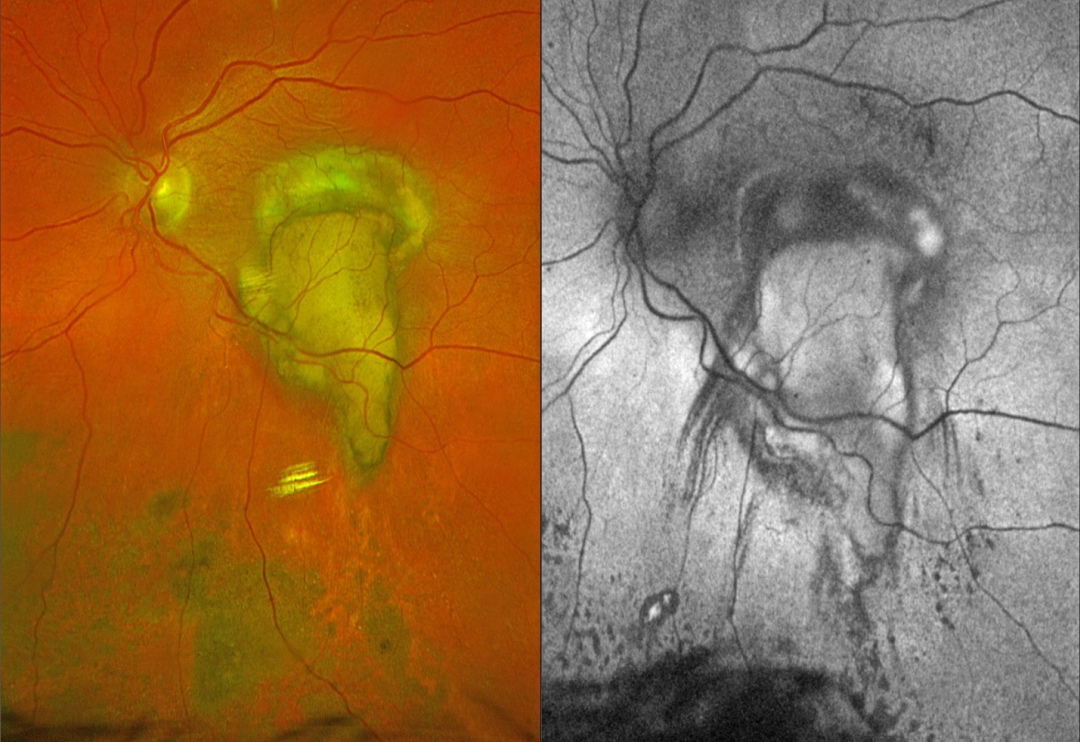
This 79YO female underwent vitrectomy for a breakthrough vitreous hemorrhage from neovascular AMD. Vision was 20/400.
Postoperative Optos color RGB imaging shows an oval fibrotic macular scar with a tongue-like projection of organized devitalized yellow blood extending inferiorly. Coarse subretinal pigment extends into the inferior midperiphery. On fundus autofluorescence (FAF), the fibrotic portion of the scar and inferior subretinal pigment are hypo-FAF, and the devitalized blood is hyper-FAF. Steaks of inferiorly radiating hypo-FAF also extend from the tongue-like yellow blood.
Learning Points:
This complex healing neovascular lesion has many signs of chronicity. There is no fresh, red blood. The remaining blood is yellow and devitalized, developing as heme products degrade. The coarse peripheral subretinal pigment is a marker of prior resolved subretinal blood and appears on OCT as subretinal hyperreflective material. Finally, the subretinal fibrosis where the breakthrough blood originated represents the usual endpoint of untreated neovascular disease, which is thankfully much less common since the advent of anti-VEGF therapy in the mid-2000s.



