

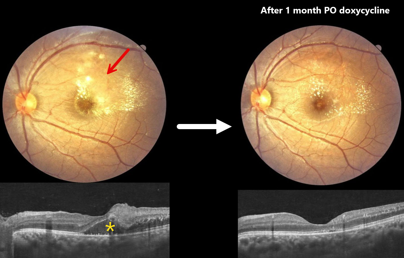
This 21YO male presented with one month of intermittent headaches and vision loss in his left eye. Vision was 20/20 in his normal OD and 20/60 OS.
Color photography of the macula shows patches of yellow-white inner retinal opacification with superior and temporal lipid. OCT shows outer nuclear layer fluid with hyperreflective dots of lipid in the outer plexiform layer. Mild vitreous cells are present.
A presumed clinical diagnosis of Bartonella neuroretinitis was made, although serology could not be performed due to cost concerns. He was started on oral doxycycline 100mg PO BID and oral corticosteroids. Two weeks later, vision was 20/40 with resolving retinal opacification and fluid (not shown). Following a full month of oral therapy, vision improved to 20/20 with decreasing lipid and no fluid.
Learning Points:
The diagnosis for a unilateral swollen nerve is extensive. However, it becomes much smaller when associated with acute inner retinal ischemia and later lipid exudation within Henle’s layer (neuroretinitis), including cat scratch (Bartonella) and syphilis. When no underlying cause is found, the entity is called Leber’s idiopathic stellate neuroretinitis.



