

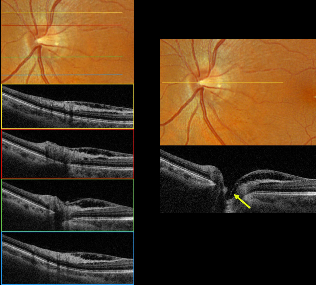
This 55YOF was referred for asymptomatic macular changes in her left eye. Vision was 20/30 bilaterally. The right eye was normal.
Optos color RGB imaging shows a normal appearing optic nerve with a small central cup. Very subtle inner retinal telangiectasia is seen inferotemporal to the disc. Triton swept-source OCT shows nasal macular inner retinoschisis, which extends into the central depression of the optic cup, but with no definite pit. This inner retinal schisis was variably present along the temporal disc margin, extending into the nasal macula. Fluorescein angiography was normal without leakage (not pictured).
Learning Points:
An optic nerve pit is a rare (about 0.1% prevalence) congenital anomaly thought to be caused by incomplete closure of the optic fissure during gestation. Pits are most often located within or along the inferior-temporal disc margin and usually appear as a gray depression. While most optic pits are asymptomatic, they can cause vision loss by forcing fluid into the inner retina, outer retina, and, ultimately, the subretinal space (optic pit maculopathy).
No optic pit retinoschisis (NOPIR) is a rare entity that causes pit-like findings in the absence of a pit or severe glaucomatous cupping (Fujimoto et al, Ophthalmology Retina 2023;7:811-818). As with optic pit maculopathy, eyes with good vision should be observed. Persistent vision loss from foveal involvement can be managed with vitrectomy, peeling of the internal limiting membrane, and fluid-gas exchange.



