

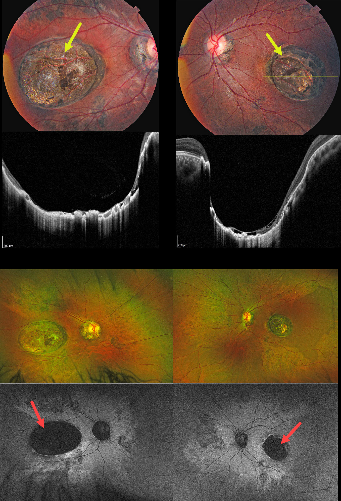
This 23YO female was referred for bilateral retinal findings. She gave a history of poor VA since early school years. Both her father and paternal grandfather had a history of vision loss since childhood. Vision was 20/60 OD and 20/80 OS. Anterior segments and motility were normal bilaterally.
Color photography shows fairly symmetrical variably pigmented colobomatous scars extending from the central maculas inferotemporally. There are patches of increased subretinal pigment along with hyperpigmented peripapillary scarring. OCT scanning through the macular lesions shows marked posterior bowing of the globe, with variable atrophy of the retina, RPE, and choroid, and hyperreflective disorganization. Variable retinoschisis is noted in the temporal aspect of the left macular lesion. On Optos fundus autofluorescence, the macular lesions and peripapillary scarring are hypo-FAF. Elsewhere, each posterior pole is hyper-FAF extending outwards from the nerves into the midperipheries. The patches of hyperpigmentation are hypo-FAF.
Genetic testing identified a heterozygous non-coding duplication upstream of the PRDM13 gene on chromosome 6q16.2, confirming the diagnosis of North Carolina macular dystrophy (NCMD).
Our patient was referred for genetic counseling and advised on visual rehabilitation options for near tasks.
Learning Points:
NCMD was originally described as a progressive autosomal dominant disorder in a pedigree from Western North Carolina (Lefler et al, AJO 1971;71:224-230). Contrary to initial expectations, the findings are stable and include symmetrical yellow spots (not drusen), macular lesions, fovea plana, and torpedo maculopathy. Vision is good unless macular neovascularization develops.
The characteristic macular lesions have variably been described as “colobomatous lesion or atrophic scar “ (Lefler 1971), “deep atrophic defects in the retina with probable loss of the choriocapillaris (Frank 1974), “staphylomatous” (Gass 1987), “crater-like or staphylomatous” (Small 1989), “macular caldera…deep chorioretinal excavations not involving the sclera” (Khurana 2009), “staphylomas” (Agarwal 2013), and “colobomatous-like lesion” (Small 2019).
The term NCMD is misleading. The findings are not limited to the macula (Green et al IOVS 2021;62(7);16), as evidenced by the pigmentary and FAF changes noted in our patient. The disease is not limited to families from North Carolina; it has been described in many subsequent pedigrees across many continents. NCMD is caused by mutations in the MCDR1 (chromosome 6) and MCDR3 (chromosome 5) genes, which encode the PRDM13 protein, a master switch controlling macular development. It has therefore been suggested that this entity be renamed simply as MCDR until the phenotypes and genotypes are better characterized (Small et al, JAMA Ophthalmology 2016;134;355-356).



