

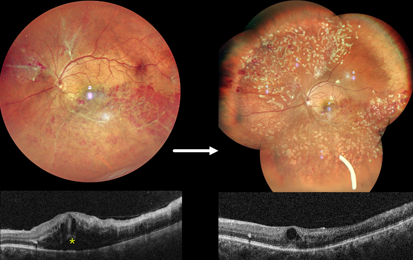
This healthy 32YO male presented on 4/23/24 with 2 months of decreased vision in his left eye. Family history was negative. A prior workup by a prior doctor was negative, including protein electrophoresis and homocysteine levels. He failed a short trial of oral steroids. Vision was 20/20 in his healthy right eye and counting fingers in his left eye.
Fundus photography shows an ischemic inferotemporal branch retinal vein occlusion with moderate, mostly peripheral retinal hemorrhages. The superotemporal retinal vein is diffusely beaded, and an apparent independent ischemic BRVO is seen superonasally, with probable preretinal blood. Marked macular edema is seen on OCT. Unfortunately, fluorescein angiography is not available.
An Ozurdex injection was given for macular edema, and two sessions of panretinal photocoagulation were performed for proliferative disease. On 6/3/24, vision improved to 20/120, and the macular edema was improving.



