

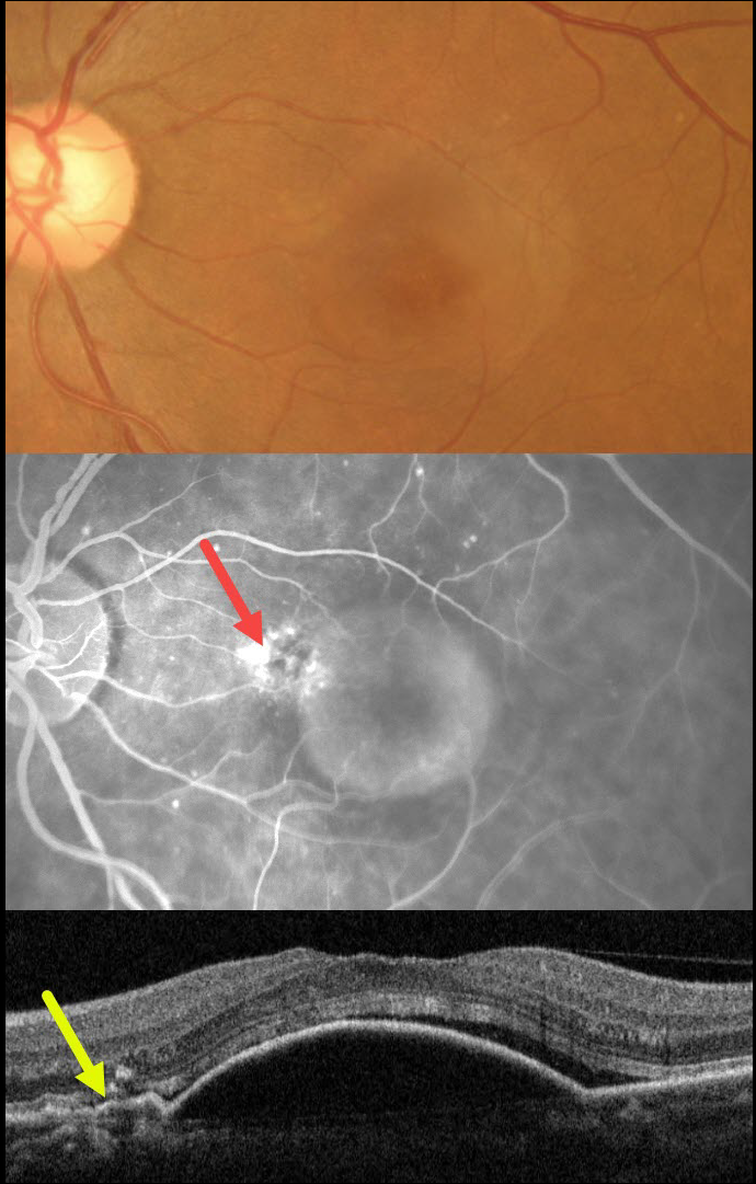
This 62YO female presented with 3 weeks of vision loss in her left eye. Vision was 20/30.
Color imaging shows a large central macular retinal pigment epithelial detachment (PED). Fluorescein angiography shows a somewhat unevenly filling PED, with staining vs leakage along its superonasal edge. OCT scanning shows a large PED, shallow overlying temporal subretinal fluid, and shallow irregular elevation of the RPE nasally indicative of a type 1 macular neovascularization (MNV).
The uneven angiographic pooling is also suggestive of an occult MNV. Anti-VEGF therapy was advised.
This case was submitted by Asma Samsudeen and Ashish Sharma.
Learning Points:
Originally described by Gass (Retina 1984;4:205-220), a notched PED is a sign of an occult MNV. The notch represents a type 1 MNV, which tethers the overlying RPE from the surrounding more highly elevated PED. Hydrostatic leakage from the MNV may contribute to the larger PED.
Although now considered a classic clinical and angiographic finding, in 1984 this was evidently not the case, as the reviewers for this article advised against publication. Based solely on who the contributing author was, the journal’s editor overruled his reviewers, published Dr. Gass’ manuscript, and the rest was literally history!



