

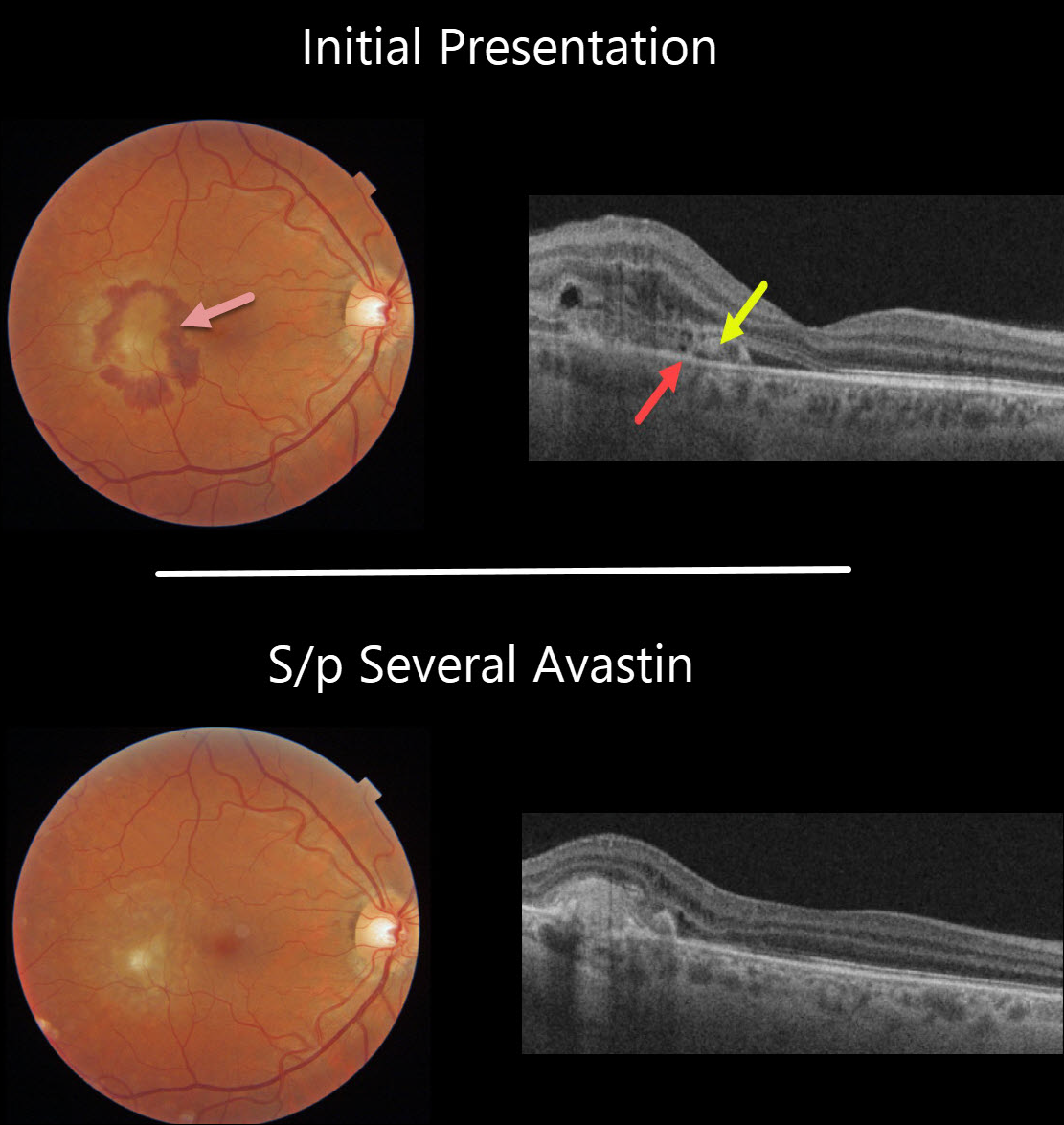
This 44YO male presented with a symptomatic macular neovascularization (MNV) from ocular histoplasmosis. Vision was 20/40.
Subretinal blood surrounds the MNV in the temporal macula. On OCT, the MNV is located above the RPE, indicating a type 2 lesion. Some retinal thickening is noted above the MNV.
The blood and fluid resolved completely following several additional monthly Avastin injections.
Learning Points:
There are three types of MNV: type 1 MNV is below the RPE, type 2 is between the RPE and outer segments, and type 3 is intraretinal.



