

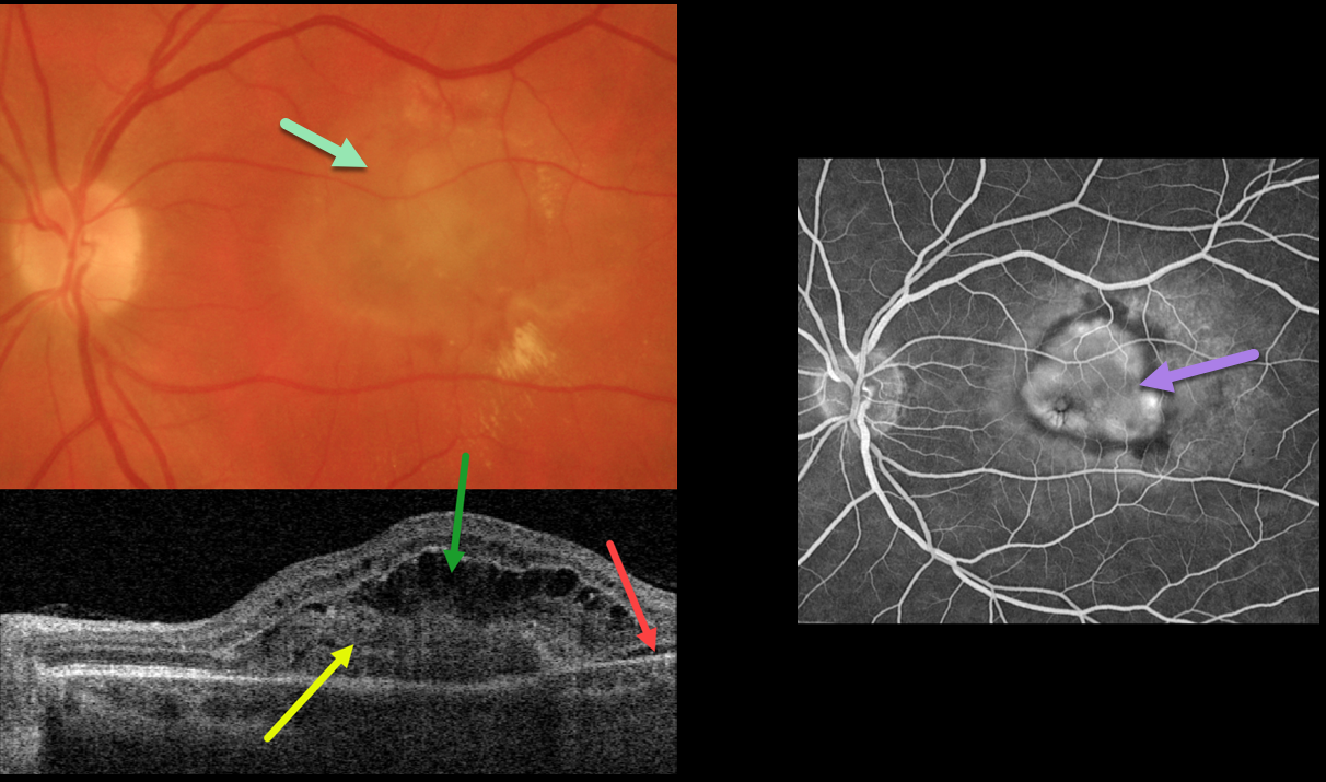
This 54YO female presented with recent vision loss in her left eye. Vision was 20/400 from an ocular histoplasmosis (OHS) related macular neovascularization (MNV).
Optos color imaging shows opaque macular fluid with a few dots of peripheral blood. Multifocal peripheral OHS scars were noted (not shown).
Swept-source OCT shows a type 2 MNV (located above the RPE), overlying outer retinal fluid and trace temporal subretinal fluid.
Fluorescein angiography shows a well-defined subfoveal lesion. Anti-VEGF therapy was recommended.
Learning Points:
Ocular histoplasmosis is caused by the Histoplasmosis capsulatum fungus which is commonly found in soil contaminated by bird or bat droppings.
It is seen worldwide, but in the United States is most often seen in the Ohio-Mississippi River valley. Most people in endemic areas inhale the organism’s spores early in life with only a few percent developing asymptomatic chorioretinal scars.
A small percentage of these will develop MNV later in life. Treatment is similar to wet AMD, with anti-VEGF therapy usually being the first line therapy. Unlike AMD, these patients often do not require ongoing injections.
Photodynamic therapy can help minimize the treatment burden, and thermal laser is still a good option for more peripheral macular lesions.



