

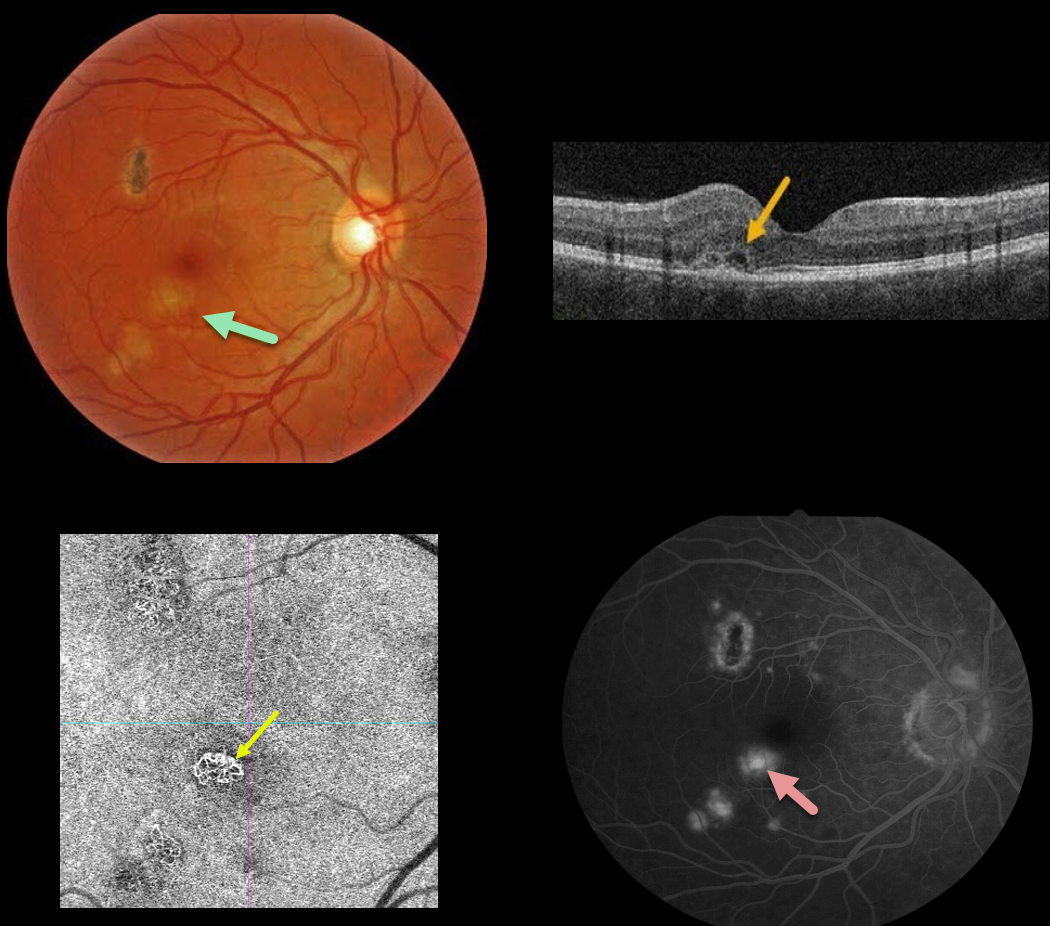
This 30YO male presented with 20/25 vision in his right eye from an ocular histoplasmosis-related macular neovascularization (MNV). Some fluid and blood were associated with a macular histoplasmosis scar in the inferotemporal fovea.
A type 2 MNV, located above the RPE, was noted on OCT. The MNV was best imaged on OCT angiography, with macular leakage noted on fluorescein angiography.
Anti-VEGF therapy was recommended.
Learning Points:
Ocular histoplasmosis is caused by the fungus Histoplasma capsulatum, which is commonly found in soil contaminated by bird or bat droppings. It is found worldwide, but in the United States, it is most common in the Ohio-Mississippi River valleys.
The vast majority of people in endemic areas inhale the organism’s spores early in life, and only a few percent will develop asymptomatic chorioretinal scars.
A small percentage of these will develop MNV later in life. Treatment is similar to wet AMD, with anti-VEGF therapy usually being the first-line therapy. Unlike AMD, these patients often do not require ongoing injections.
Photodynamic therapy can help minimize treatment burden, and thermal laser therapy remains a good option for more peripheral macular lesions.



