

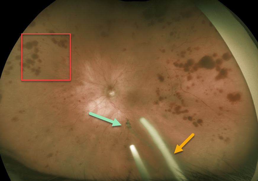
This 86YO patient was receiving Ozurdex (dexamethasone 0.7%) injections every 8 weeks in her left eye for macular edema from an inferior hemiretinal vein occlusion.
She subsequently developed ocular ischemic syndrome (OIS) like changes with scattered peripheral large outer retinal hemorrhages. Inferiorly, more recent Ozurdex implants are noted, along with another ghost-like remnant of an older implant. Although the Ozurdex implants are supposed to completely degrade, these remnants may persist beyond 1 year (see Kim et al Retina 2020;40;2226-2231).
Learning Points:
The retinal hemorrhages in ocular ischemic syndrome (OIS) are unique. Retinal vascular disorders like diabetic retinopathy, retinal vein occlusions, and hypertensive retinopathy usually have flame- and dot-shaped inner retinal hemorrhages concentrated around the posterior pole.
Our patient’s findings are classic for OIS, with hemorrhages in the deeper peripheral retina. The retinal periphery is literally the end of the line for the retinal arterial circulation. In an eye receiving less blood supply due to carotid occlusive disease, the more proximal retina has first dibs on the available oxygen. There are penetrating capillaries that dive radially from the nerve fiber and ganglion cell layers into the deeper retina. The deep vascular complex that supplies the inner and outer plexiform layers is thus furthest downstream, and these endothelial cells are likely damaged in OIS. Blood cells leaking into this space accumulate, causing the large outer blot to hemorrhage.
These patients need to have a carotid Doppler and ultrasound and be referred for medical or surgical intervention if they have a critical stenosis. They may also develop anterior or posterior segment neovascularization requiring photocoagulation and/or anti-VEGF injections. Iris neovascularization can develop, leading to neovascular glaucoma. Hypotony can also occur.



