

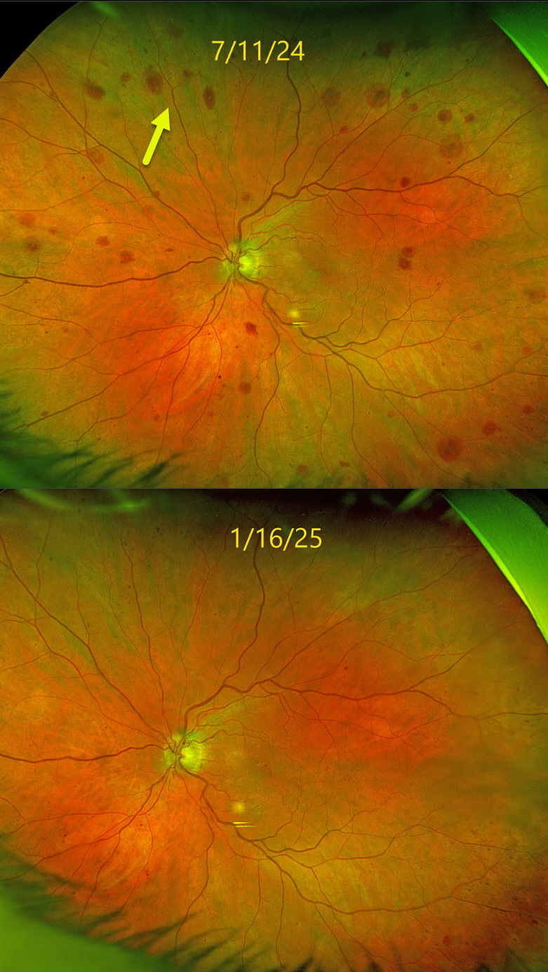
This 83YO male was referred for asymptomatic changes in his left fundus. Vision was 20/30 in his healthy OD and 20/100 OS.
Optos color RG imaging shows scattered deep, variably round retinal hemorrhages extending from the peripheral macula into the midperiphery. Carotid Doppler testing revealed a critical stenosis of the left internal carotid artery, and he was emergently referred to his cardiologist. The findings dramatically improved within months of carotid surgery.
Learning Points:
The retinal hemorrhages in the ocular ischemic syndrome (OIS) are unique. Retinal vascular disorders like diabetic retinopathy, retinal vein occlusions, and hypertensive retinopathy usually have flame- and dot-shaped inner retinal hemorrhages concentrated around the posterior pole.
Our patient’s findings are classic for OIS, with hemorrhages in the deeper peripheral retina. The retinal periphery is literally the end of the line for the retinal arterial circulation. In an eye receiving less blood supply due to carotid occlusive disease, the more proximal retina has first dibs on the available oxygen. There are penetrating capillaries that dive radially from the nerve fiber and ganglion cell layers into the deeper retina. The deep vascular complex that supplies the inner and outer plexiform layers is thus furthest downstream, and these endothelial cells are likely damaged by OIS. Blood cells leaking into this space accumulate, giving the large outer blot hemorrhages.



