

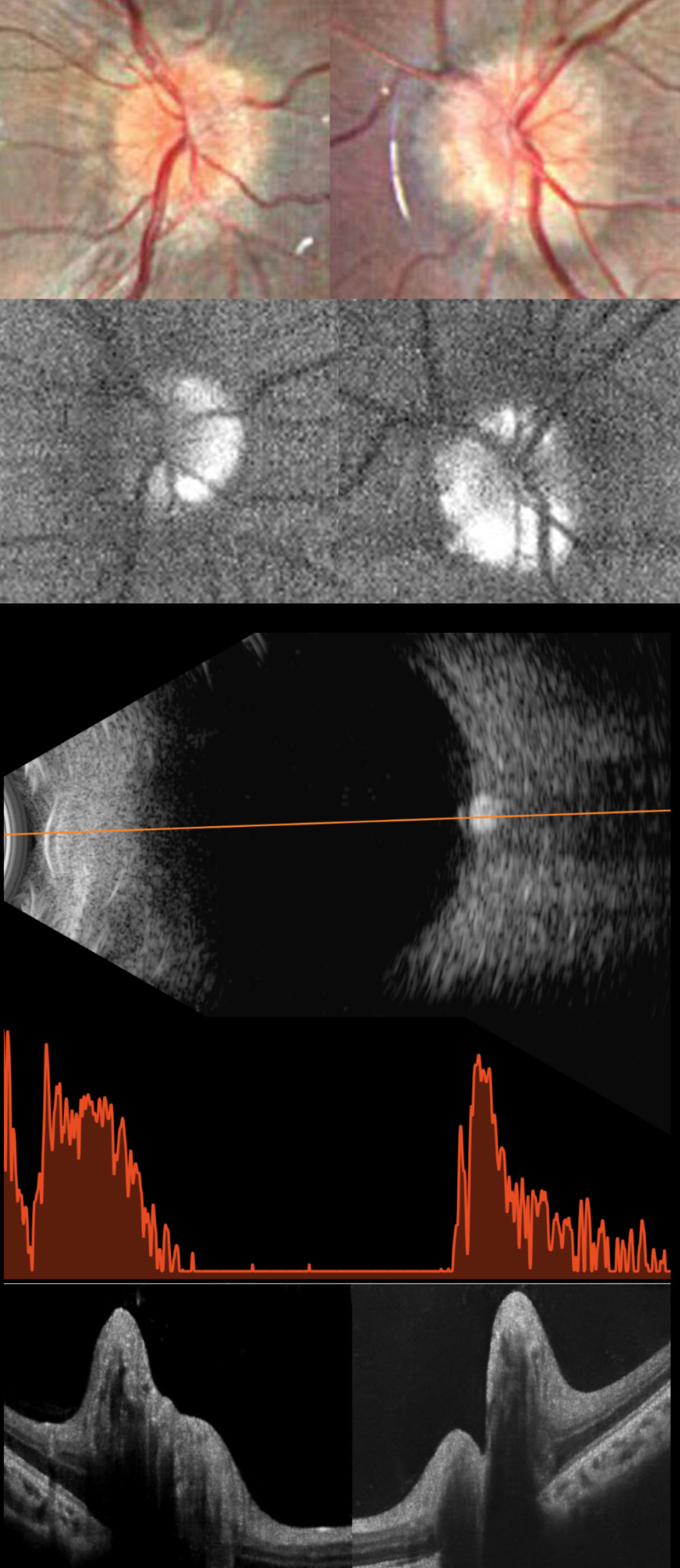
This healthy 11YO girl was diagnosed elsewhere 2 months earlier with papilledema and precocious puberty, which was being treated with oral acetazolamide. Workup included a normal brain MRI and lumbar puncture with normal opening pressure and CSF analysis. Vision in our office was 20/20 OU.
Fundus photography shows pseudopapilledema with extensive optic disc drusen (ODD). On fundus autofluorescence (FAF), the drusen are hyper-FAF. On B-scan ultrasonography, the discs are markedly hyperreflective, indicating calcification. OCT scanning shows marked, irregular disc thickening due to drusen.
Learning Points:
Optic disc drusen (ODD) are refractile calcific concretions that form anterior to the lamina cribrosa. They are thought to represent extruded, extracellular, calcified mitochondria and may be inherited as an autosomal dominant trait.
Porphyrins, organic molecules composed of four interconnected pyrrole subunits, are present within mitochondria. When these compounds break down, they become autofluorescent, which is likely why drusen autofluoresce. Porphyrins are also part of the heme molecule in hemoglobin, which explains why devitalized blood autofluoresces (Bloom and Spaide, Retinal Cases 2022;16:401-402).
ODD can be confused with true disc swelling, as occurred in our patient, which led to an erroneous diagnosis of papilledema. In our experience, the pathognomonic B-scan ultrasonographic disc calcification is rarely seen in patients this young, but is extremely helpful in establishing the diagnosis when present. We find FAF to be a more reliable test when the ODD are not obvious clinically. After speaking with her neurologist, the acetazolamide was discontinued, and observation was recommended.



