

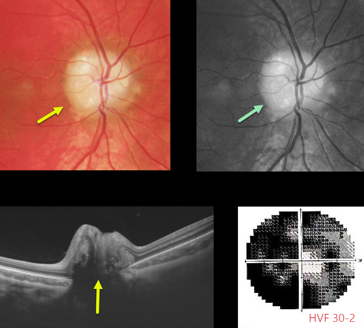
Our patient is a 15YO boy with quite pronounced bilateral optic nerve drusen visible clinically, as well as on OCT (only right eye pictured). ODD will often hyperautofluoresce on fundus autofluorescence (FAF).
Although central vision was good, he complained of increasing peripheral vision loss due to drusen compressing the surrounding nerve fibers.
Learning Points:
Optic disc drusen (ODD) are refractile calcific concretions that form anterior to the lamina cribrosa. They are thought to be extruded, extracellular, calcified mitochondria and may be inherited as an autosomal dominant trait.
It is often difficult to differentiate ODD from true optic nerve edema. This is often the dilemma when seeing a child with a possible swollen nerve.
We have found ultrasonography to be unhelpful, since ODD don’t calcify until later in life.
B-scan OCT imaging is often unhelpful, although newer swept-source machines can directly visualize drusen. The RNFL will be variably thinned with ODD and thickened with true disc edema.
Fluorescein angiography (FA) is probably the best way to differentiate the two, since a swollen nerve will show late leakage, whereas ODD will not leak and often shows staining of the drusen.
Unfortunately, there is no proven treatment, although some have tried pressure-lowering medications for neuroprotection.



