

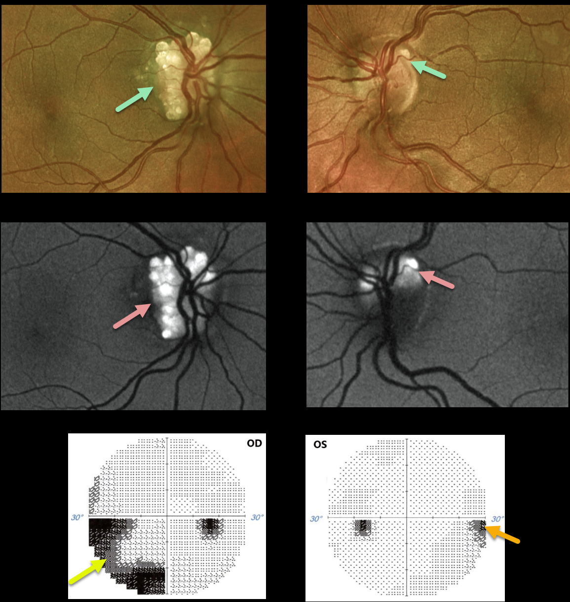
This 38YO female presented with bilateral optic disc drusen (ODD). Vision was 20/80 OD and 20/40 OS.
The drusen are markedly hyper-autofluorescent. The retinal nerve fiber is thinned, particularly in the right eye, due to the more extensive ODD. Humphrey visual fields show significant visual field defects inferonasally OD and mild nasal depression OS. Observation was recommended.
Learning Points:
Optic disc drusen (ODD) are refractile calcific concretions that form anterior to the lamina cribrosa. They are thought to be extruded, extracellular, calcified mitochondria and may be inherited as an autosomal dominant trait.
Porphyrins, organic molecules composed of four interconnected pyrrole subunits, are present within mitochondria. When these compounds break down, they become autofluorescent, which is likely why drusen autofluoresce.
Porphyrins are also part of the heme molecule in hemoglobin, accounting for why devitalized blood autofluoresces.



