

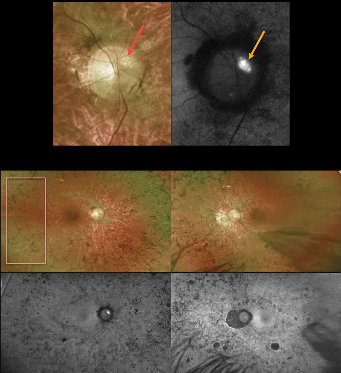
This 43YO male with a known history of retinitis pigmentosa (RP) was referred for further evaluation. He noted a gradual worsening of vision bilaterally. Vision was 20/80 OD and 20/200 OS.
Optos color imaging of the right optic nerve shows several large optic disc drusen (ODD) that markedly hyperautofluoresce. Ultrawide-field imaging shows diffuse, classic bilateral RP scarring.
Learning Points:
ODD are refractile calcific concretions that form anterior to the lamina cribrosa. They are thought to be extruded, extracellular, calcified mitochondria and may be inherited as an autosomal dominant trait. They are present in about 3% of RP patients (Russell et al, Eye 2022;36:2213).
Most patients are asymptomatic, but some can develop progressive visual field defects or central vision loss. There is no treatment for the drusen themselves, although symptomatic macular neovascularization can be treated with thermal laser, photodynamic therapy, or anti-VEGF injections.



