

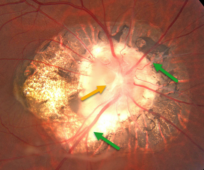
This 43YO female presented for a routine examination. There was a history of pathological myopia. Vision was 20/70 OD and 20/60 OS.
Optos color imaging shows peripapillary scarring with a morning-glory optic disc, including a large disc, a central glial tissue area, and numerous straight, radiating vessels. Observation was recommended.
Learning Points:
Morning glory syndrome is typically unilateral and shares some similarities with optic disc coloboma and juxtapapillary staphyloma. This abnormality gets its name from its striking resemblance to the Morning Glory flower. Visual prognosis is usually poor. The condition can be associated with serous macular detachment. Neuroimaging should be considered to rule out transsphenoidal encephalocele and intracranial carotid artery dysgenesis.
For a great review on the pathogenesis and treatment of maculopathy associated with cavitary optic disc anomalies, see Jain and Johnson, AJO 2014;158:423-435.



