

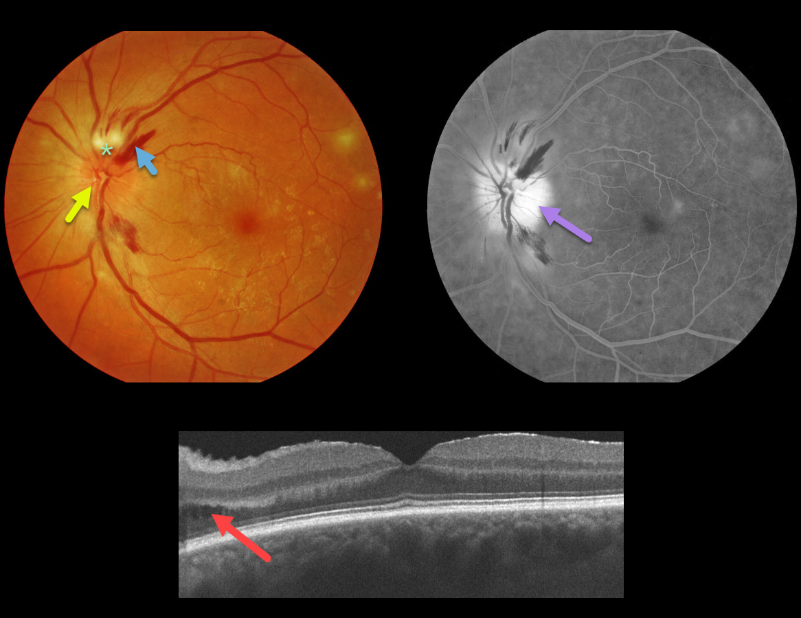
This 66YO diabetic male presented with an asymptomatic unilateral swollen nerve, peripapillary hemorrhages, and nerve fiber layer ischemia. Vision was 20/25, and there was no afferent pupillary defect (APD).
OCT scanning shows some mild cystic fluid in the peripapillary inner and outer nuclear layer. Fluorescein angiography shows diffuse optic nerve leakage.
Blood pressure was normal. He denied exposure to deer ticks or cat scratches, and the MRI was normal. The funduscopic findings spontaneously normalized within a few months.
Learning Points:
Diabetic papillopathy, often a diagnosis of exclusion, is most commonly seen in type 2 diabetics. It has a good visual prognosis with observation alone. A small physiologic cup may predispose to this condition (see Regillo et al, Arch Ophthlamol 1995;113:889-895).
Unlike ischemic optic neuropathy, vision is usually quite good, there is usually no APD, the nerve swelling is more diffuse than sectorial, and there is no altitudinal field defect.
The differential for unilateral acute optic nerve swelling is broad, including diabetes, ischemic (anterior ischemic optic neuropathy), and inflammatory causes (optic neuritis, Bartonella, syphilis, sarcoidosis).
Papillitis must be differentiated from papilledema, which is bilateral optic nerve swelling caused by elevated intracranial pressure.
We have been burned more than once by unilateral nerve edema being the only presenting sign of severe hypertension. Although nerve swelling is usually bilateral in hypertensive retinopathy, we always check blood pressure in all patients presenting with unilateral or bilateral optic nerve swelling.



