

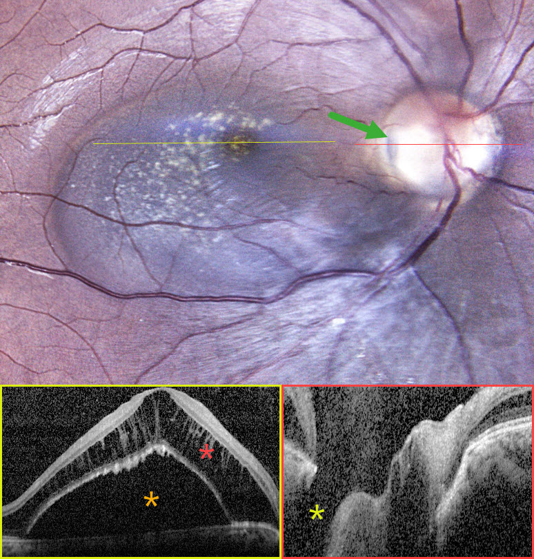
This healthy 24YO female presented with asymptomatic retinal changes in her right eye, which was 20/20 one year earlier. Vision was 20/80 OD and 20/20 in her normal OS.
Fundus photography shows a serous macular detachment connected to a yellow-grey optic nerve pit along the temporal edge of the disc. OCT scanning shows an elevated serous detachment with marked overlying cystic schisis in the outer nuclear layer. The pit is noted on the OCT.
Learning Points:
An optic nerve pit is a rare (about 0.1% prevalence) congenital anomaly thought to be caused by incomplete closure of the optic fissure during gestation. Pits are most often located within or along the inferior-temporal disc margin and usually appear as a gray depression. While most optic pits are asymptomatic, they can cause vision loss by forcing fluid into the inner retina, outer retina, and, ultimately, the subretinal space (optic pit maculopathy). The source of the fluid remains a subject of ongoing debate, with opinions ranging from the vitreous to the subarachnoid space.
Given the lack of symptoms, continued observation was recommended for our patient.



