

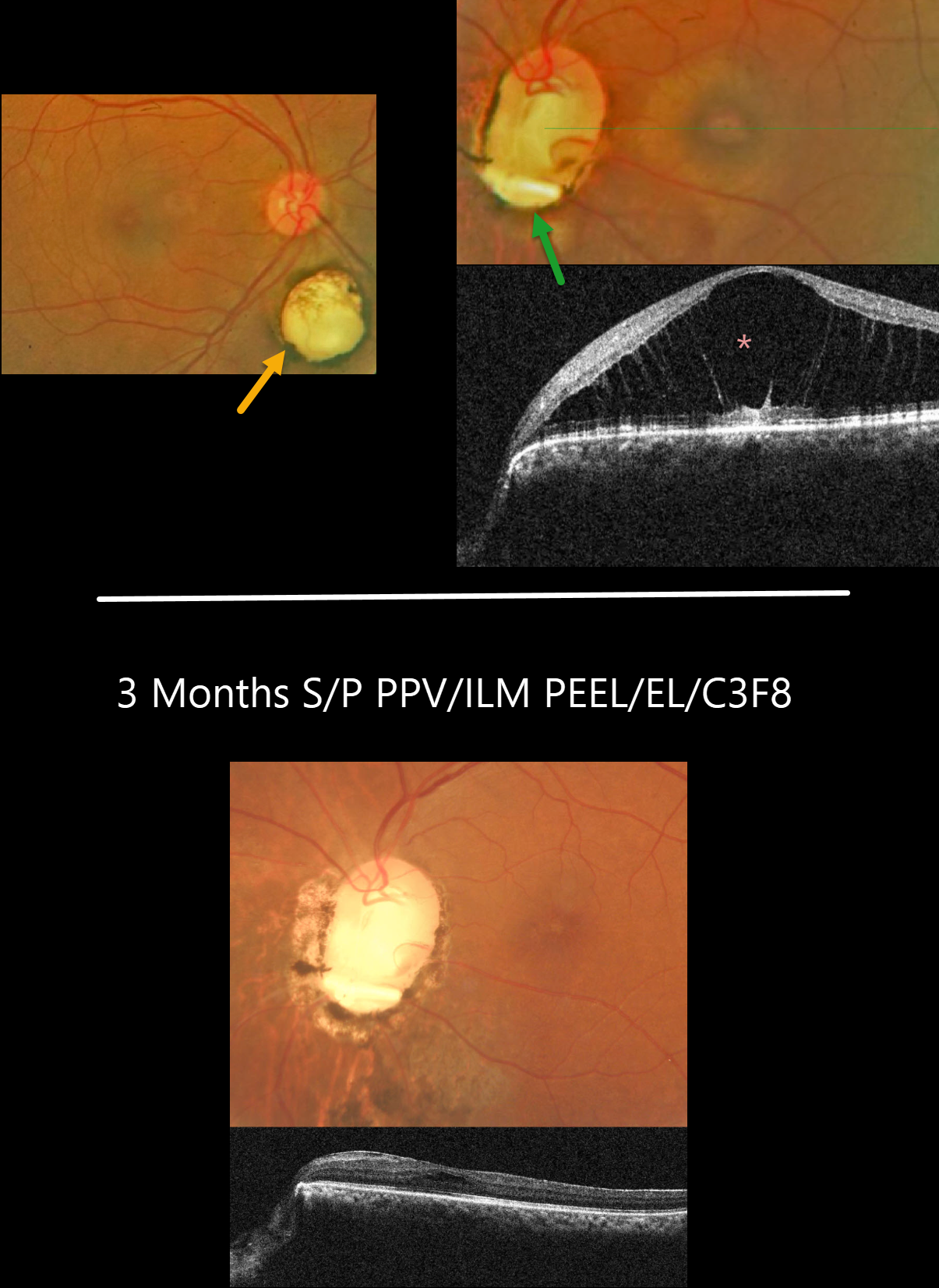
This 51YO female presented with 1 month of blurred vision in her left eye. There was no past ocular history. Vision was 20/30 OD and 20/80 OS. Anterior segments were normal.
Fundus photography of her right eye shows a small choroidal coloboma inferior to the nerve. The left nerve is anomalous with an inferior coloboma and optic pit. The central macula was elevated, with severe foveoschisis noted on OCT.
Vitrectomy with ILM peeling, endolaser along the disc margin, and C3F8 was performed, followed by some additional thermal laser around the nerve postoperatively.
Three months following vitrectomy, vision improved to 20/40 with complete flattening of the macular schisis.
Learning Points:
An optic nerve coloboma can be unilateral or bilateral and is caused by incomplete closure of the embryonic fissure. It has a pathogenesis similar to optic nerve pits, and our patient’s left nerve shows features of both.
While most optic pits are asymptomatic, they can cause vision loss by forcing fluid into the inner retina, outer retina, and, ultimately, the subretinal space (optic pit maculopathy).
The source of the fluid remains a point of ongoing debate, with the fluid originating from either the vitreous or subarachnoid space.
A host of treatments for optic pit maculopathy have been suggested over the years. However, a recent meta-analysis suggested that a plain-vanilla vitrectomy without gas tamponade is as successful as other procedures (Zheng et al., Ophthalmology Retina 2020;4:289-299).



