

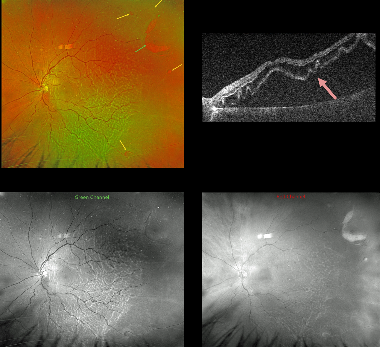
This 61YO male presented with a several-week history of severe vision loss in his left eye. Vision was hand motion.
Optos color RG imaging shows a rhegmatogenous retinal detachment (RRD) with corrugated outer retinal folds. A large horseshoe retinal tear is seen superotemporally, with numerous smaller breaks noted.
The outer retinal folds are best seen with the green channel compared with the red channel. Triton swept-source OCT shows how these findings involve just the outer retina.
Vitrectomy surgery was scheduled.
Learning Points:
Hydration of outer retinal folds is one of the key findings to help differentiate a RRD from exudative and traction retinal detachments. The cause for these folds remains uncertain, but they may form as the result of the hydration of glycosaminoglycans in the interphotoreceptor matrix (Dalvin et al, Retinal Cases & Brief Reports 2020;14:355-359).



