

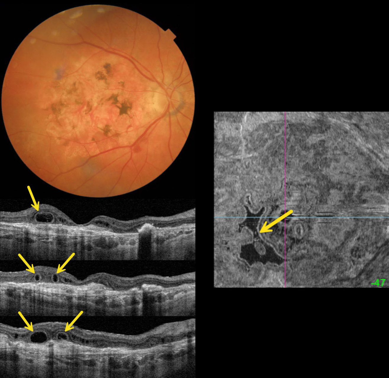
This 89YO female is receiving ongoing intravitreal Eylea in her 20/40 left eye for neovascular AMD. She has a stable 20/400 atrophic disciform scar in her right eye.
Triton color imaging shows a variably pigmented flat disciform scar. Swept-source OCT shows variable outer retinal atrophy overlying a hyperreflective inactive macular neovascularization (MNV). A complex pattern of outer retinal tubulation (ORT) is seen on numerous B-scans. En face OCT shows another view of these meandering tubular structures.
Learning Points:
ORT is often noted overlying inactive MNV and should not be confused with exudative fluid or cysts, which lack a hyperreflective border. The outer hyperreflective band likely represents inner segment mitochondria undergoing fission and translocation toward the nucleus (Litts et al, Retina 2018;38:445-461). ORT, initially described by Zweifel et al (Arch Ophthalmol 2009;127:1596-1602), is a neurodegenerative condition of the photoreceptors and Muller cells associated with atrophy affecting the outer retina and retinal pigment epithelium, including advanced AMD and inherited retinal diseases.



