

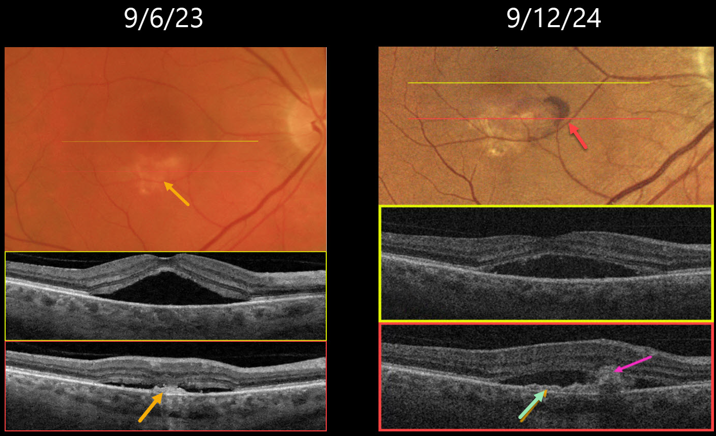
This 64YO male presented on 9/6/23 with several weeks of vision loss in his right eye. Vision was 20/40 OD.
Triton color imaging shows a faint round foveal serous detachment. Swept-source OCT confirms a classic idiopathic central serous chorioretinopathy (ICSC) foveal detachment. There is a small hyperreflective placoid elevation of the RPE along the inferior edge of the detachment corresponding to a yellow-white area of subretinal pigmentation. Due to significant symptoms, he elected for early photodynamic therapy (PDT), which successfully resolved the serous fluid two months later with 20/30 vision (not shown).
He did well until he returned on 9/12/24 with recurrent blurred vision OD, which had decreased slightly to 20/40. There is new subretinal blood within the region of the persistent placoid RPE elevation inferiorly. OCT shows recurrent serous foveal fluid centrally. There is an enlargement of the shallow inferior RPE elevation adjacent to the subretinal blood. Monthly intravitreal Avastin therapy was started.
Learning Points:
Initially described by Pang and Freund, pachychoroid neovasculopathy is a variant of type 1 (below the RPE) macular neovascularization (MNV) found as part of the pachychoroid spectrum, including ICSC and pachychoroid pigment epitheliopathy (Retina 2015;35:1-9). About one-third of chronic ICSC eyes with flat irregular RPE detachments will harbor an MNV on OCT angiography (Bousquet et al, Retina 2018;38:629-639), which is probably a form of pachychoroid neovascularization (Hwang et al, Retina 2020;40:1724-1733).
In retrospect, our patient most likely had a pachychoroid neovascularization, which initially responded to PDT. When he returned one year later with a definite active MNV, we elected to start anti-VEGF monotherapy. He was unfortunately immediately lost to follow-up.



