

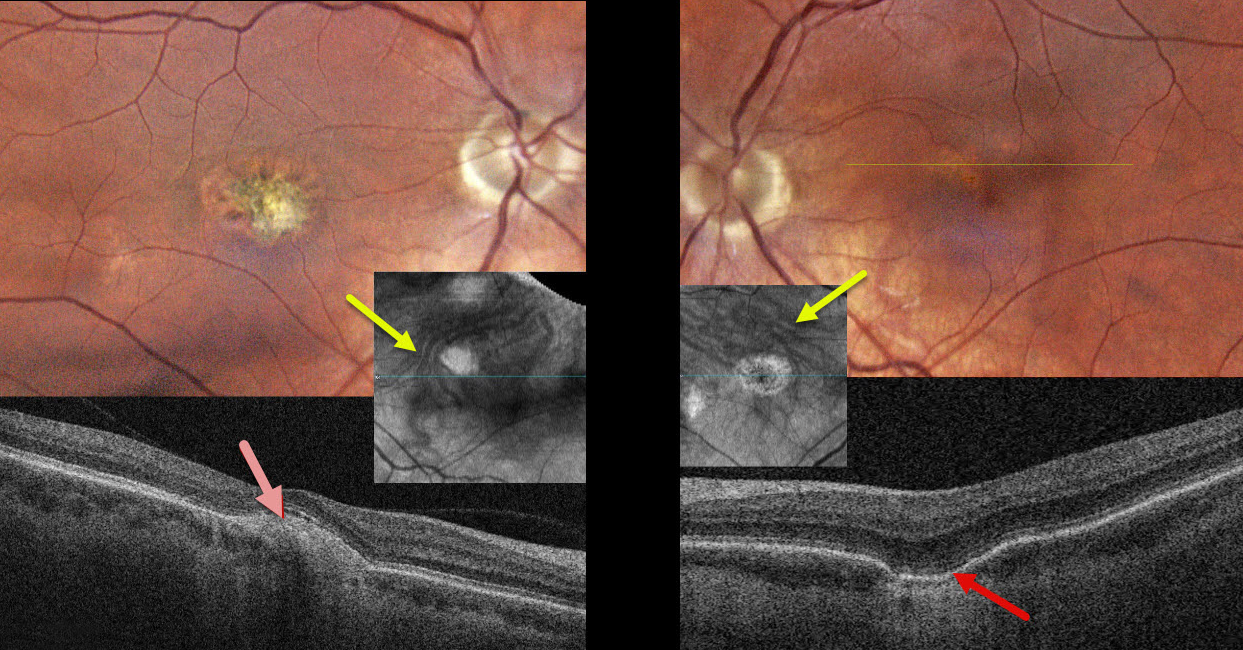
This healthy 39YO female gave a history 18 years earlier of prior anti-VEGF therapy OD for ocular histoplasmosis. She was visually asymptomatic, and vision was 20/40 OD and 20/30 OS.
Color photography of her left eye shows a patchy area of pigment loss in the superonasal fovea. OCT scanning through this area shows a choroidal excavation. The right macula has an overall mostly depigmented area of scarring, which appears as a mound of subretinal hyperreflectivity on OCT. Choroidal en face OCT shows dilated pachyvessels OU that cross the horizontal meridian (vortex anastomoses). There were no areas of peripheral chorioretinal scarring in either eye. Observation was recommended.
Learning Points:
Pachychoroid, meaning “thick” choroid, is secondary to large, dilated outer choroidal vessels. Increased oncotic pressure from these dilated vessels can cause RPE dysfunction, serous detachment, and macular neovascularization (MNV). Pachychoroid disease spectrum is a growing list of findings, including idiopathic central serous chorioretinopathy (ICSC), pachychoroid neovascularization, polypoidal choroidal vasculopathy, pachychoroid pigment epitheliopathy, pachydrusen, peripapillary pachychoroid syndrome, choroidal excavation, and pachyvitelliform maculopathy.
Our guess is that our patient had active ICSC when she was treated for her ‘ocular histoplasmosis.’ The lesion in the right macula is atypical for ICSC. But the other findings, including dilated choroidal vessels with vortex anastomoses and a choroidal excavation in her left eye, argue for pachychoroid as her unifying diagnosis.



