

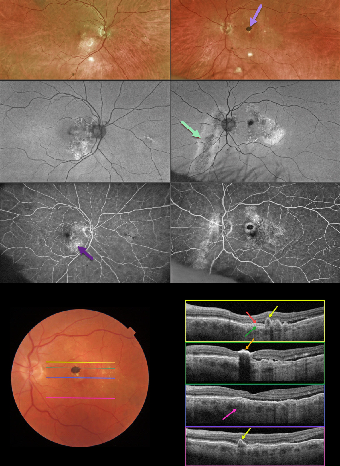
This 78YO male was referred for asymptomatic fundoscopic changes. Vision was 20/25 OD and 20/30 OS. He had a history of laser treatment in his left eye decades earlier.
Optos imaging shows bilateral areas of pigmentary loss in each macula, extending inferiorly. A hyperpigmented laser scar is noted in the left superior macula.
Fundus autofluorescence (FAF) shows variable hypo- and hyper-FAF, along with a prominent gutter extending from the left nerve inferonasally.
Fundus fluorescein angiography (FFA) shows window defects within these areas, with blockage from the hyperpigmented laser scar OS.
Swept-source OCT shows numerous findings. Scanning superior to the laser scar shows several choroidal polyps. A shallow elevation of the RPE exposes the underlying Bruch’s membrane. Scanning through the laser scar shows intense hyperreflectivity with posterior shadowing. Scanning through the macular center shows temporal subretinal fluid, along with a thickened choroid. Finally, scanning in the inferior macula shows an additional polyp.
Learning Points:
This case has an overwhelming amount of features from the pachychoroid spectrum, which includes idiopathic central serous retinopathy (ICSC), pachychoroid neovasculopathy, polypoidal choroidal vasculopathy, pachychoroid pigment epitheliopathy, peripapillary pachychoroid syndrome, and choroidal excavation.
The thermal laser treatment many years ago may have been for pachychoroid neovasculopathy, a recently described phenotype (see Pang and Freund, Retina 2015;35:1-9) found in the pachychoroid spectrum. These lesions are particularly common in patients with ICSC, and often remain silent as a flat, irregular RPE elevation on OCT without exudative complications.
Our patient likely had further progression of their pachychoroid disease, resulting in multiple polyps, which are found in polypoidal choroidal vasculopathy (PCV). PCV is a variant of type 1 macular neovascularization, also in the pachychoroid spectrum. PCV is likely distinct from age-related macular degeneration, occurring in a different population (more common in African Americans and Asians), with a relative lack of drusen, and the presence of a thicker choroid.
In addition to the findings above, the patient had a large gutter in his left eye, a common finding in ICSC. Gutters form from pathologies with chronic leakage. Conditions that form gutters other than ICSC include choroidal hemangiomas, choroidal nevi, and uveal melanoma.
For more information on the pachychoroid spectrum, see Cheung et al, Eye 2019;33:14-33 for a great pachychoroid disease review.



