

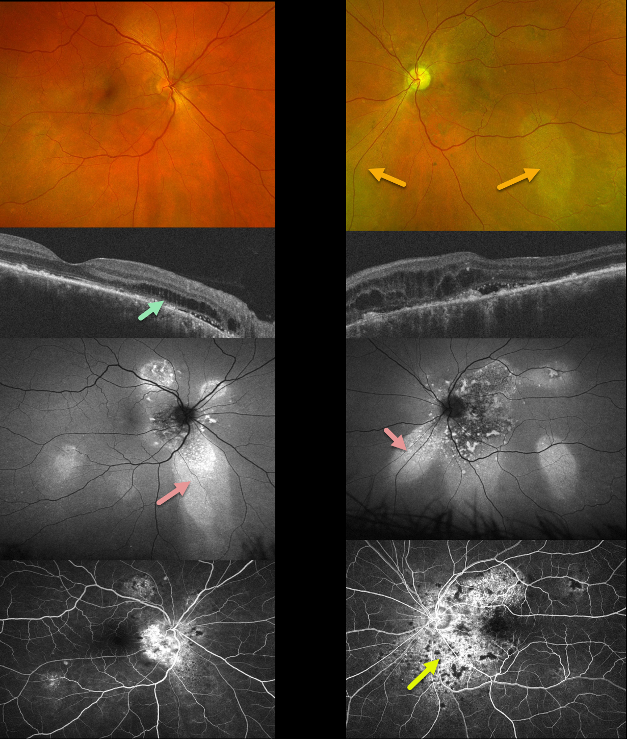
This 51YO male has been followed for years with self-limited episodes of idiopathic central serous retinopathy (ICSC).
Optos color imaging of both eyes shows regions of inferior pigmentary loss, most pronounced in the OS. These findings are much more dramatic on fundus autofluorescence. Swept-source OCT shows variable bilateral loss of the outer retinal bands. Outer retinal fluid with trace subretinal fluid is seen centrally and nasally OS. Fluorescein angiography shows variable staining.
Learning Points:
This patient has numerous features of the pachychoroid spectrum, including idiopathic central serous retinopathy (ICSC), pachychoroid neovasculopathy, polypoidal choroidal vasculopathy, pachychoroid pigment epitheliopathy, peripapillary pachychoroid syndrome, and choroidal excavation.
Our patient’s current findings are dominated by the peripapillary pachychoroid syndrome. Originally described by Phashukkuwatana et al (Retina 2018;1652-1667), findings include peripapillary choroidal thickening associated with nasal macular subretinal and/or intraretinal fluid. Most patients also have chorioretinal folds.
The fluid waxes and wanes, and is responsive to photodynamic therapy if treatment is needed for persistent symptomatic foveal fluid (Iovino et al, Eye 2022;36:716-723). Our patient is being observed for now.



