

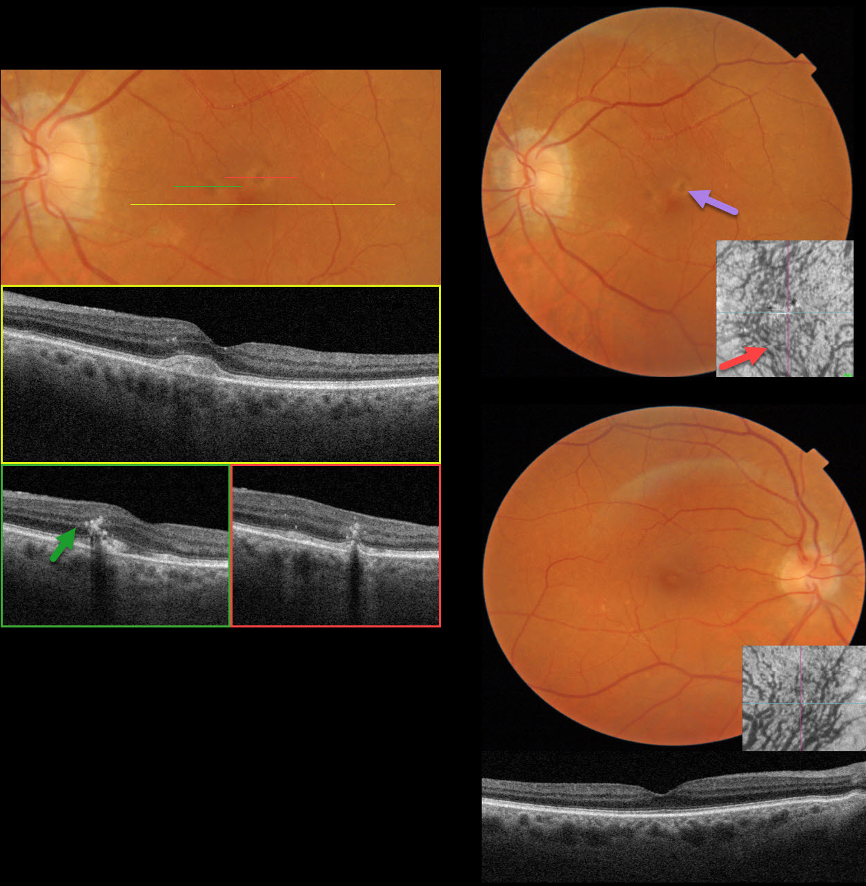
This 81YO female was referred for asymptomatic retinal findings. Vision was 20/30 OD and 20/30 OS.
Triton color imaging OS shows several yellowish subretinal pigment clumps in the superior fovea. Swept-source OCT through the central macula shows an acquired vitelliform lesion (AVL). Scans through each of the pigment clumps show coarse intraretinal hyperreflective foci. Some ectopic pachydrusen are noted in the inferotemporal right macula. OCT shows large outer choroidal vessels, and en face OCT shows the entire macula to be drained by the inferior vortex system with dilated vessels. The choroidal vessels are also dilated OS.
Learning Points:
Pachychoroid, meaning “thick” choroid, is secondary to large, dilated outer choroidal vessels. Increased oncotic or mechanical pressure from these dilated vessels can cause RPE dysfunction, serous detachment, and macular neovascularization (MNV). Our patient has pachychoroid findings despite having a ‘normal’ choroidal thickness on B-scan OCT. The choroidal thickness depends on numerous factors, including the patient’s age and refractive error. A high myope’s choroid should be quite thin, but if it appears as a ‘normal’ thickness, it is thick for that eye. We also often see pachychoroid disease in elderly patients whose choroid is abnormally thickened for their age, but appears ‘normally’ thick when not accounting for this.
Pachychoroid disease spectrum includes central serous chorioretinopathy, pachychoroid neovascularization, polypoidal choroidal vasculopathy, pachychoroid pigment epitheliopathy, pachydrusen, peripapillary pachychoroid syndrome, and focal choroidal excavation (Cheung et al, Eye 2025;39:819-834 ). Hilely et al recently described an additional entity, pachyvitelliform maculopathy (Br J Ophthalmol 2024;108:753-759), defined by AVL associated with pachychoroid features. The authors hypothesize that the thickened choroid causes RPE dysfunction, resulting in the accumulation of undigested photoreceptor outer segments with AVL formation.
We believe a better term for these disorders is venous overload choroidopathy, described by Spaide et al (Progress in Retinal and Eye Research 2022;86:100972). This hypothesis suggests scleral venous outflow as a unifying cause for this ever-expanding spectrum.



