

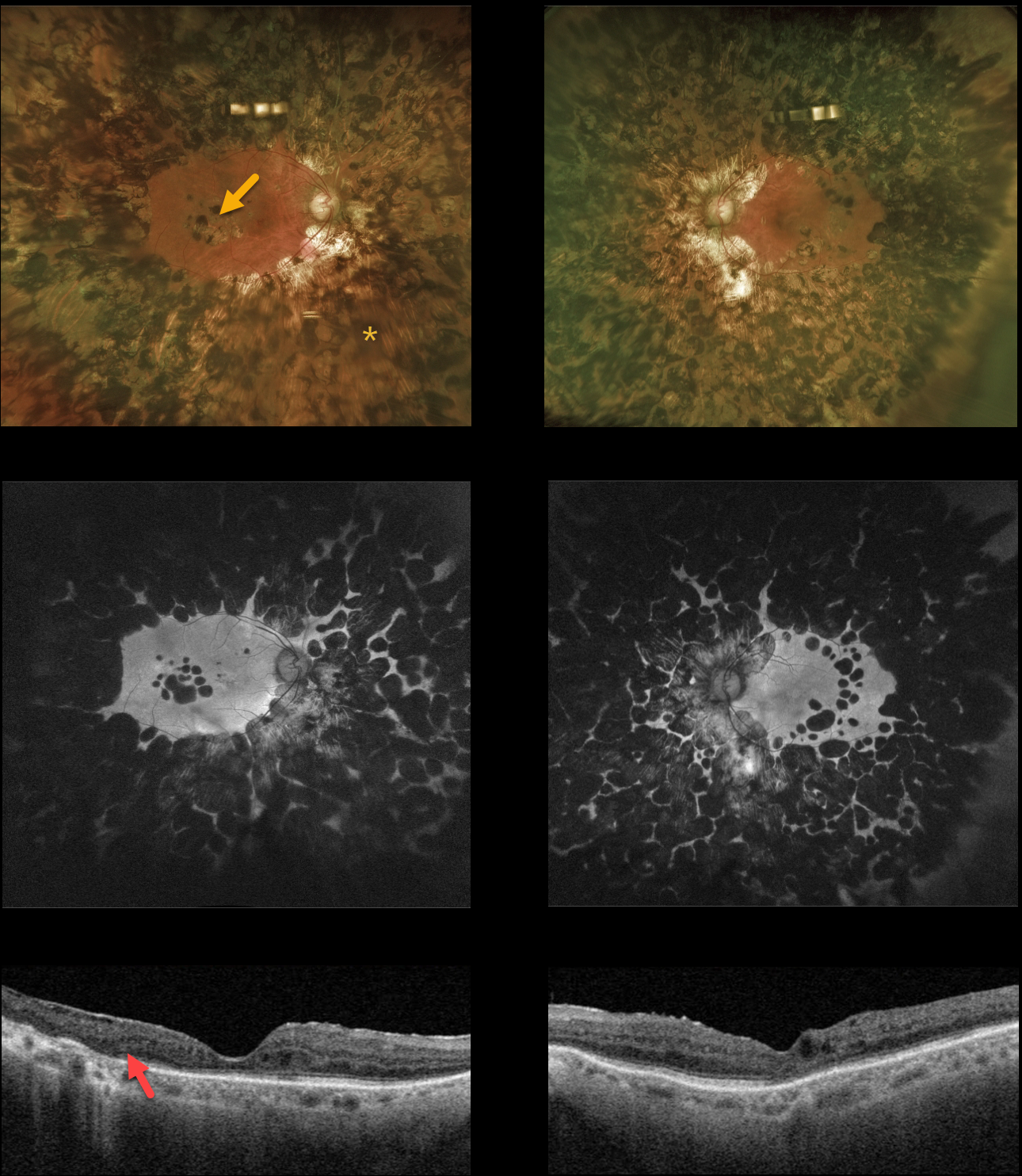
This 80YO diabetic female gave a history 0f prior retinal lasers elsewhere about 20 years earlier. Vision was 20/30 OD and 20/80 OS.
Optos color and fundus autofluorescent imaging show heavy bilateral panretinal photocoagulation (PRP) and macular laser scarring. Swept source OCT shows variable disorganization of the retinal layers and outer retinal atrophy. Visual fields (not shown) are severely constricted due to the ‘iatrogenic’ retinitis pigmentosa caused by the posterior and confluent laser scarring.
Learning Points:
Although possibly appropriate before the advent of modern vitrectomy surgery and anti-VEGF therapy, this degree of laser should never be seen in today’s patients. Laser should begin well outside the arcades and away from the optic nerve, extending as anteriorly as possible with the 200 to 500 micron-sized retinal burns spaced about one burn-width apart.



