

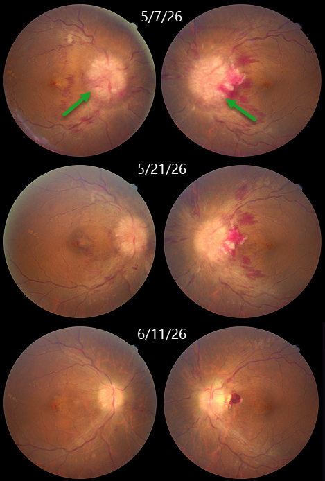
This 44YO obese female presented with several days of decreased vision bilaterally. Vision was counting fingers OU. Fundus photography shows severe bilateral disc swelling.
MRI of the brain and orbit was suggestive of partial empty sella with prominent perioptic CSF spaces and no intracranial mass lesions. All these were suggestive of idiopathic intracranial hypertension (IIH). She was referred to neurology, where a spinal tap established the diagnosis of IIH, and she was started on acetazolamide 1000 mg qday. One month later, vision improved to 20/20 OU with marked improvement in the papilledema.
Learning Points:
IIH, formerly known as pseudotumor cerebri, is caused by elevated intracranial pressure primarily due to decreased cerebrospinal fluid (CSF) drainage caused by elevated cerebral venous pressure. The condition is most often found in obese women of childbearing age. Presenting symptoms include headaches, transient visual obscurations, neck/back pain, and tinnitus/whooshing. Treatment includes weight loss, acetazolamide, and topiramate. Severe cases unresponsive to medical therapy, or those with transverse sinus stenosis, require surgery (CSF diversion procedures, venous sinus stenting, or optic nerve sheath fenestration).



