

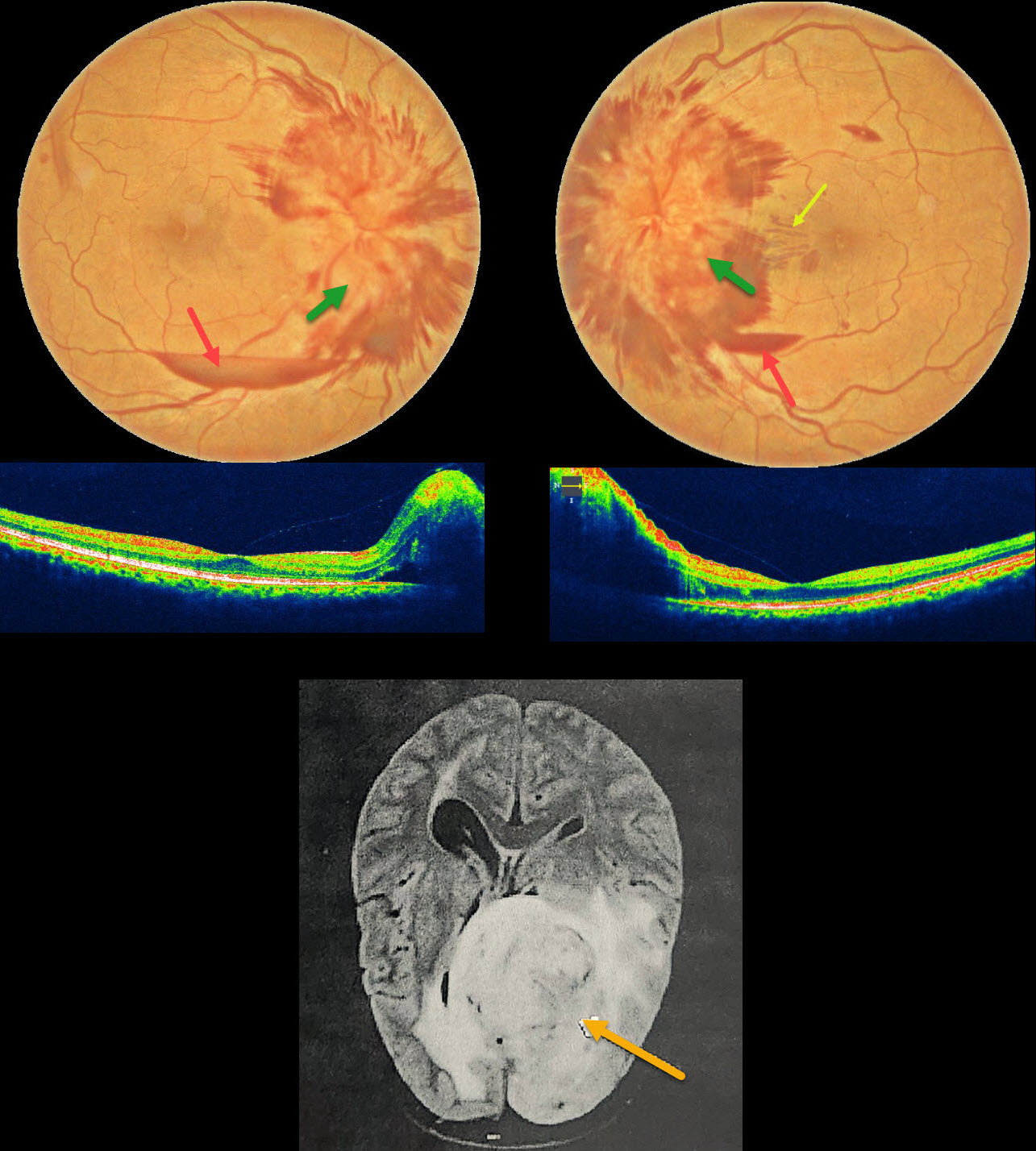
This 50YO female presented with one month of bilateral vision loss. Vision was 20/90 OD and 20/70 OS.
Fundus photography shows severe bilateral optic nerve swelling with surrounding nerve fiber layer hemorrhages. Small inferior subhyaloid hemorrhages are present bilaterally, along with a small outer plexiform layer hemorrhage in the left nasal macula.
MRI scanning showed a high-grade glioma, and she was immediately referred to neurosurgery for further management.
Learning Points:
Glioblastoma multiforme, also known as a grade IV astrocytoma, represents the most common and aggressive primary malignant brain tumor. A variety of neuro-ophthalmic signs and symptoms result from direct involvement of the visual pathway or elevated intracranial pressure, including vision loss or visual field defects, afferent pupillary defect, extraocular muscle motility abnormalities, and papilledema. Although most patients rarely survive beyond 1 year from diagnosis, the prognosis is especially poor with optic nerve involvement.



