

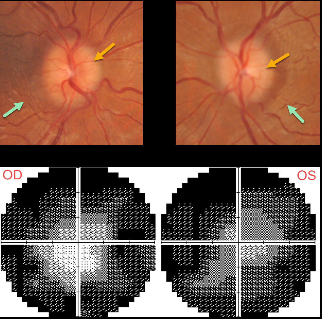
This 30YO female was diagnosed with idiopathic intracranial hypertension (pseudotumor cerebri) in 2020. Her initial lumbar puncture opening pressure was elevated at 25cm H2O.
She was lost to follow-up for about 2 years, when she presented with recurrent headaches, a ‘whooshing’ sound in her ears, and blurred vision. She was off of Topamax for about a year. Vision was 20/30 OD and 20/40 OS.
Both optic nerves are mildly edematous. The retinal nerve fiber layer (not shown) is somewhat thickened bilaterally. Despite the modest nerve edema, prominent temporal circumferential retinal folds (Paton’s folds) surround both nerves. Visual fields are severely constricted.
Learning Points:
In 1911 Paton and Holmes established that papilledema was due to elevated intracranial pressure and not inflammation as was previously thought (Brain 1911;33:389-432).
They also noted that nerve edema was associated with “a series of folds which run concentric with the edge of the disc” which they felt represented retinal folds.
Sibony and Kupersmith, correlating OCT with fundus photography, found that these folds represented a variety of structural stresses on the nerve and peripapillary tissues, including peripapillary wrinkles, inner retinal folds, choroidal folds and outer retinal folds/creases (Ophthalmology 2016;123:1397-1399).



