

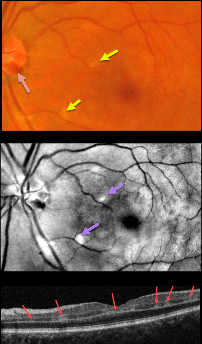
This 57YO female presented with sudden painless vision loss in her left eye. There was no significant past ocular or medical history. Vision was 20/20 in her normal right eye and 20/130 in her left eye.
Fundus photography shows several small hemorrhages along the temporal disc margin and faint patches of scattered whitish retinal opacification, which are much more pronounced in a perivenular distribution in the red-free image.
OCT B-scan shows multiple patches of focal hyperreflectivity involving the inner plexiform, inner nuclear, and outer plexiform layers.
Learning Points:
Paracentral acute middle maculopathy (PAMM) is a recently described phenotype that consists of acute mid-retinal ischemia, occurring as an idiopathic entity or in other ocular disorders, including Purtscher’s, diabetic retinopathy, retinal vascular occlusions, and acute macular neuroretinopathy.
For a great recent review, see Scharf et al, Progress in Retinal and Eye Research 2021;81;100884.
Perivenular PAMM may be the only presenting sign for an incomplete central retinal artery occlusion or mild central retinal vein occlusion (see Zhao et al, AJO 2022;234:15-19). Our patient may indeed have a very mild CRVO, given the optic nerve hemorrhages.



