

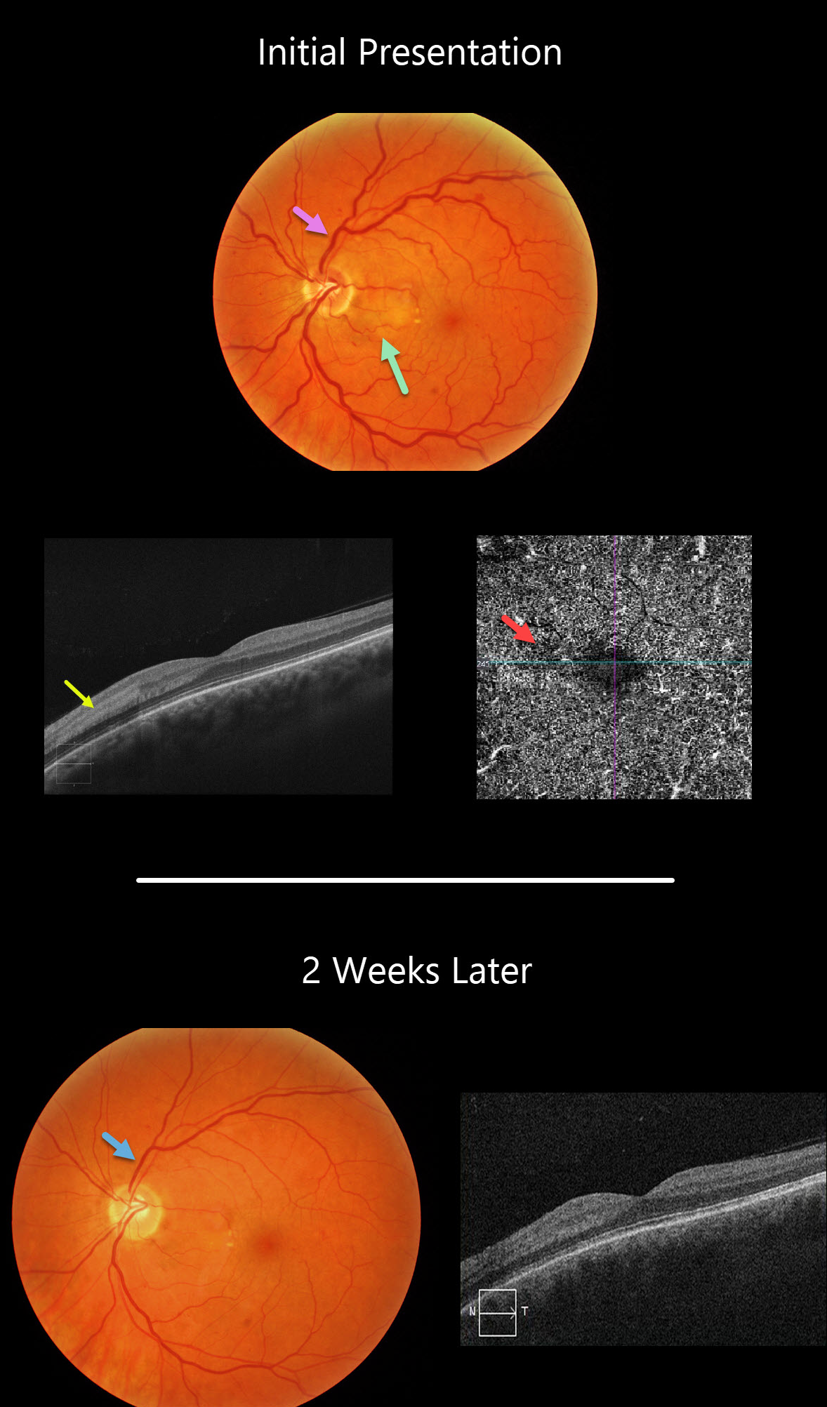
This 45YO female presented with 20/40 vision in her left eye with an acute negative scotoma temporal to her visual axis. This corresponded with a focal area of retinal whitening in the nasal macula.
This appeared as hyperreflectivity of the inner plexiform, inner nuclear, and outer plexiform layers on OCT and a relative flow void on OCT angiography.
Two weeks later, the retina and OCT findings improved, and vision was 20/30. This may have represented a mild, spontaneously resolving central retinal vein occlusion, as the initial fundus photo shows very mild, scattered retinal hemorrhages with dilated veins, and the venous caliber normalized at her exam 2 weeks later.
Learning Points:
Paracentral acute maculopathy (PAMM) is a recently described phenotype.
It consists of acute mid-retinal ischemia found as an idiopathic entity or in other ocular disorders, including Purtscher’s, diabetic retinopathy, retinal artery and retinal vein occlusions, and acute macular neuroretinopathy.



