

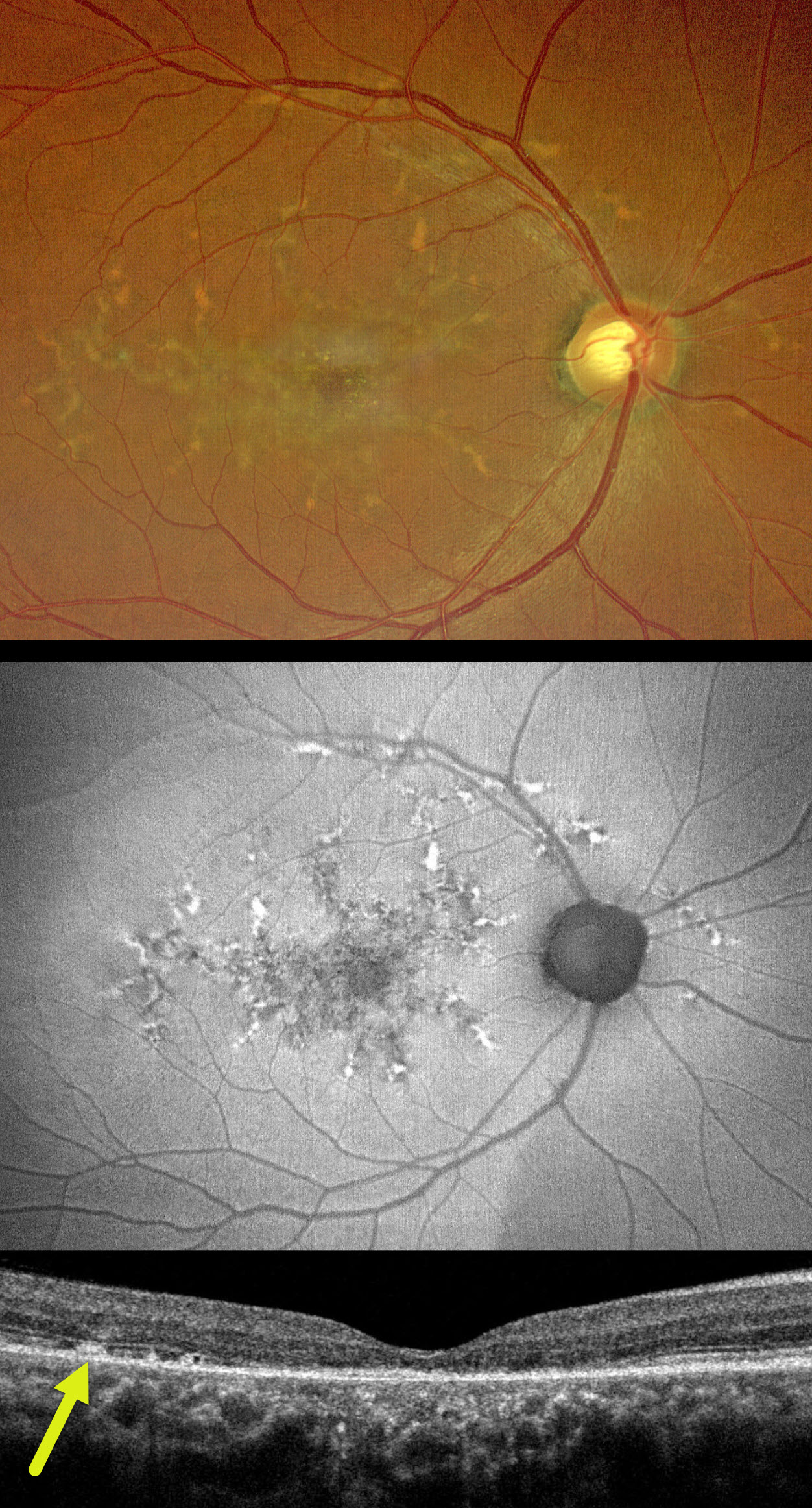
This healthy 50YO male presented with 10 months of bilateral vision loss. Family history was negative. Vision was 20/80 OU.
Color photography of his right eye shows submacular pigment and orange-yellow flecks radiating outwards from the fovea towards the peripheral macula and around the nerve. These lesions on fundus autofluorescence (FAF) are variably hyper- and hypo-FAF. OCT shows variable outer retinal loss. The subretinal flecks appear as hyperreflective deposits above the RPE. Identical findings are present in his left eye (not shown).
Learning Points:
Autosomal dominant pattern macular dystrophies present in mid-life with macular subretinal orange-yellow or pigmented deposits that accumulate in various patterns. Usually caused by mutations in the PRPH2 gene, the condition is classified by the pattern created by these deposits. However, they likely represent different phenotypes of the same disorder. The most common clinical presentation is adult-onset foveomacular vitelliform dystrophy, which presents as symmetrical, small, subfoveal, yellow-pigmented lesions. Our patient’s appearance is characteristic of butterfly-shaped pigment dystrophy. The visual prognosis is generally good, although some patients may develop vision loss due to secondary atrophy or macular neovascularization.



