

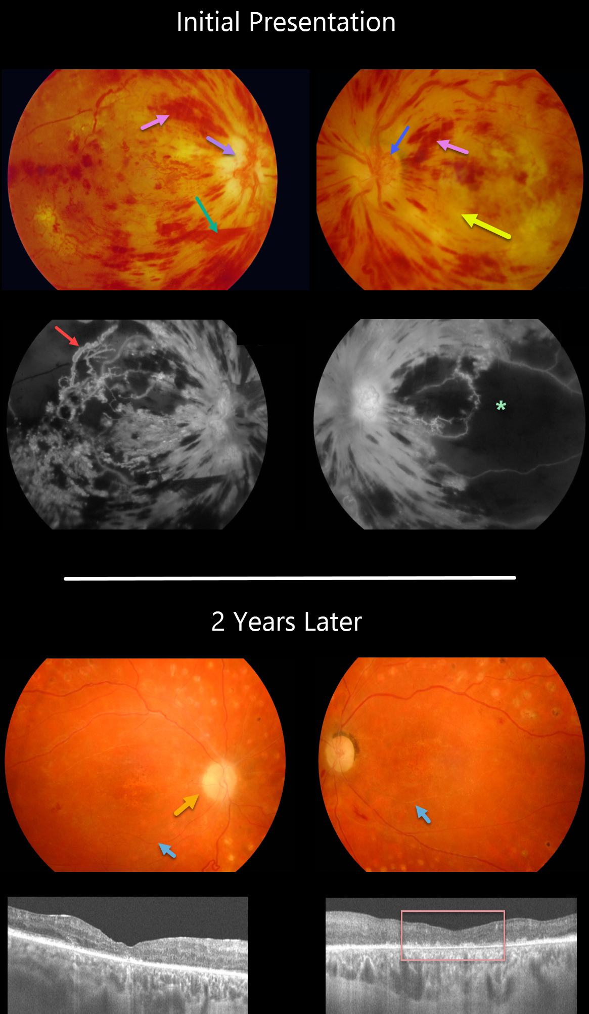
This 30YO female presented in 2008 with acute severe bilateral vision loss. She had a history of well-controlled type 1 diabetes.
There were severe bilateral retinal hemorrhages with white, ischemic maculas. A small preretinal hemorrhage was present just inferior to the right nerve. The right optic nerve was swollen. Disc neovascularization was noted in her left eye.
Fluorescein angiography showed severe macular ischemia, especially in her left eye, with near total obliteration of the normal macular vascular anatomy. Prominent staining venous beading was present in the right superotemporal macula.
Her blood pressure was severely elevated. Following emergent hypertensive control, bilateral anti-VEGF therapy was given, followed by panretinal photocoagulation.
Two years later, vision returned to 20/40 OD and 20/400 OS. The optic nerves were pale, the retinal vessels were narrowed and sheathed, and the foveas had dry pigmentary changes.
OCT scanning showed thinned, atrophic maculae with disorganized inner retinal layers (DRIL).
Learning Points:
Malignant hypertension, defined as blood pressure above 180 systolic and/or 120 diastolic, is a life-threatening medical emergency. Eye doctors are in a unique position to often diagnose this condition. Patients will often present with bilateral optic nerve swelling, flame-shaped retinal hemorrhages, and, with more chronic disease, lipid precipitates in the nasal macular outer plexiform layer.
Hypertensive retinopathy can have a similar appearance to diabetic retinopathy, and patients (like this one) can even present with signs of both.
Over time, patients with hypertensive retinopathy will develop near complete regression of all findings following systemic blood pressure treatment and control.



