

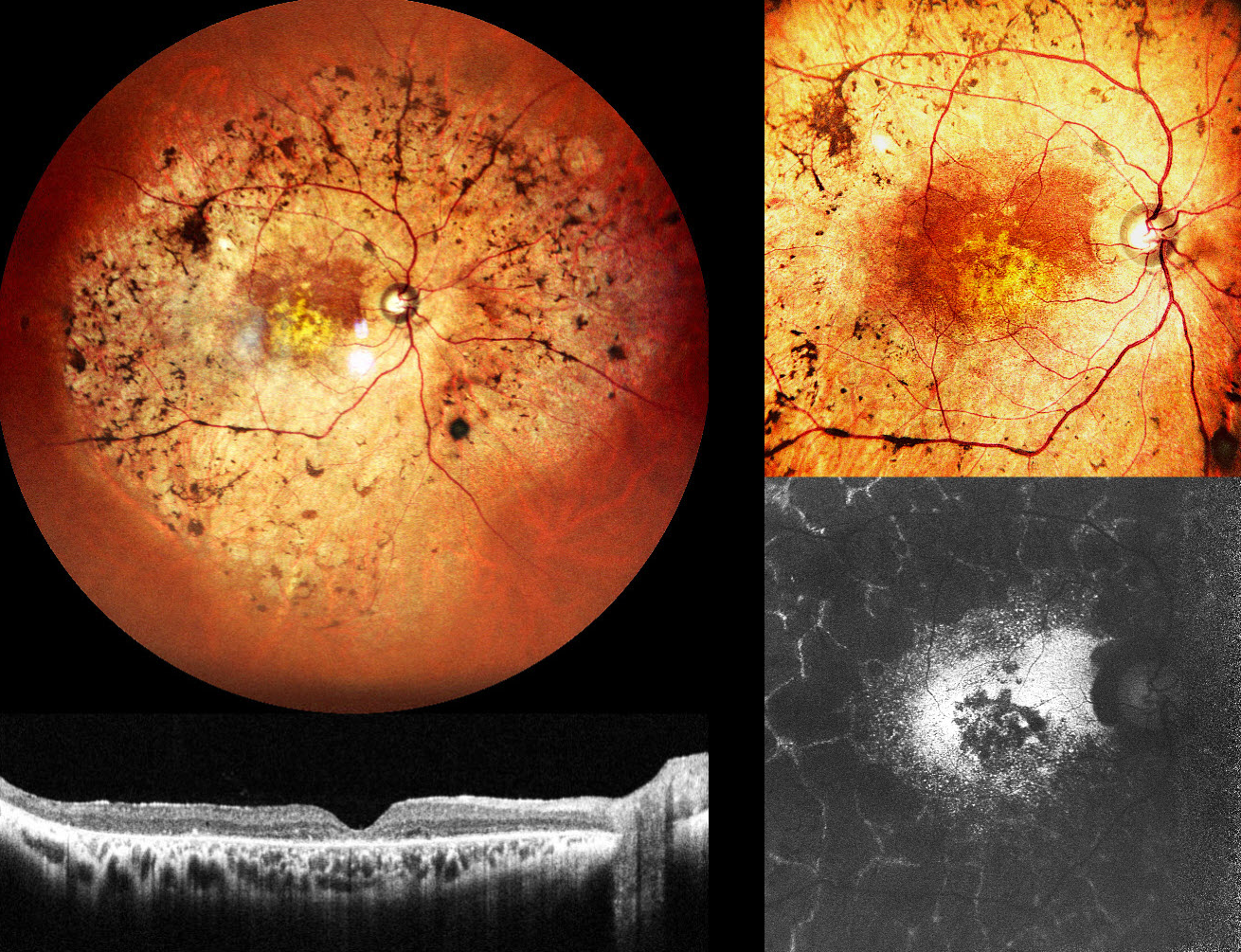
This 47YO male presented with decreased vision in both eyes for 5 years. Ocular family history was negative. Vision was 20/300 OD and 20/660 OS.
Pseudocolor SLO imaging of his right eye shows intraretinal pigment migration (bone-spicules) extending from the macula into the midperiphery. OCT shows outer retinal thinning with disorganization of the outer retinal bands. Fundus autofluorescence (FAF) shows hyper-FAF from the relatively preserved paracentral retina, more central hypo-FAF, and a peculiar peripheral pattern of interlaced hyper-FAF lines around lobular atrophic RPE and retina. Identical findings were noted in his left eye (not shown).
Learning Points:
Pericentral retinitis pigmentosa (RP) is an atypical, mild form of RP characterized by retinal degeneration and intraretinal pigment migration confined to the retinal near midperiphery. Unlike classic RP, pericentral RP tends to progress more slowly, with preserved central visual acuity and less extensive visual field loss over time. Genetically, pericentral RP is heterogeneous (Matsui et al, IOVS 2025;56:6007-6018), with causative variants identified in multiple genes, many of which are also associated with typical RP and other retinal dystrophies, indicating that pericentral RP is a phenotypic variant rather than a distinct clinical entity. Long-term prognosis for central vision is generally favorable in the absence of macular involvement, which unfortunately



