

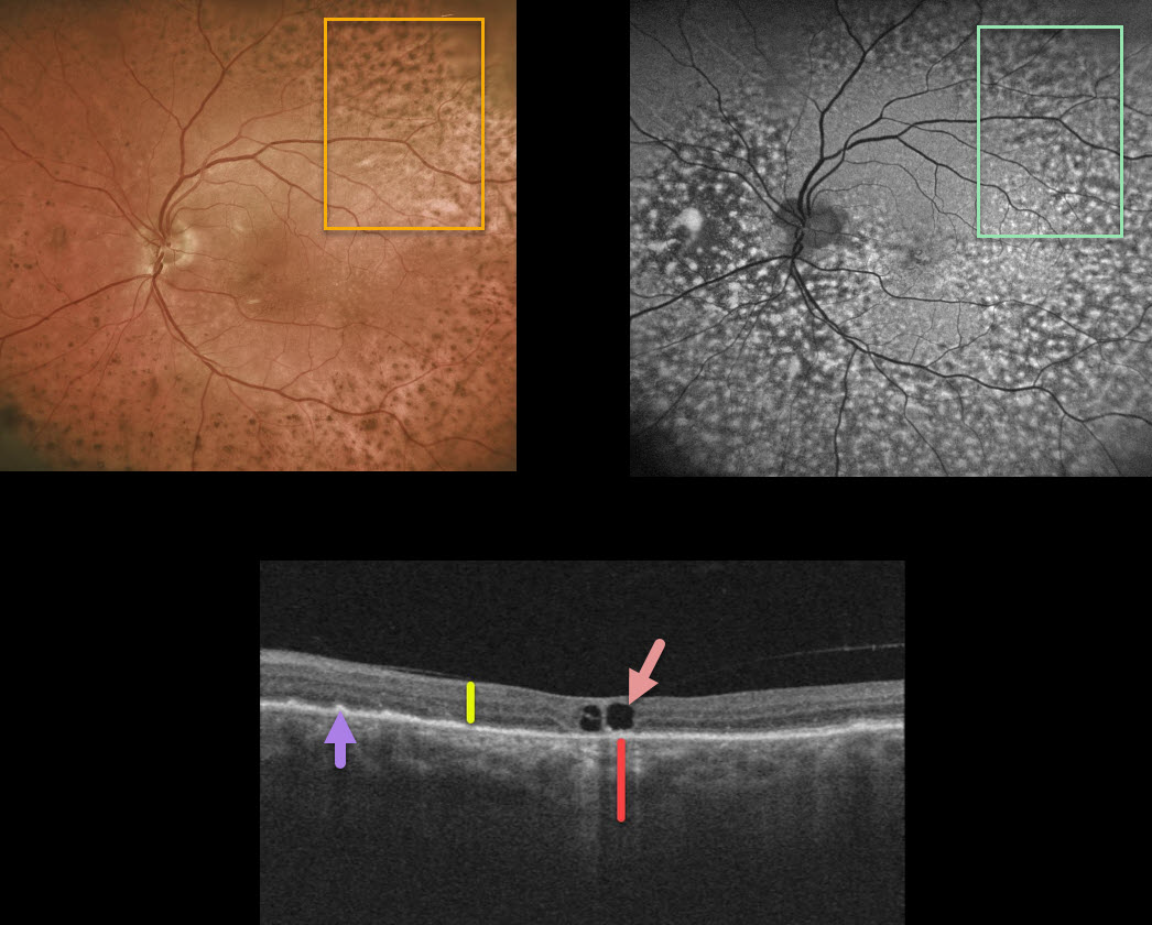
While being followed for peripapillary pachychroid syndrome (PPS) with periodic photodynamic therapy for symptomatic foveal fluid OS, this patient presented with new diffuse pigmentary deposits in the left eye only.
Fundus autofluorescence shows a dramatic pattern of hyperautofluorescent and hypofluorescent spots scattered throughout the fundus. These findings, although unilateral, look very similar to those in the paraneoplastic condition, bilateral diffuse uveal melanocytic proliferation (BDUMP).
Macular OCT scanning shows a diffusely thickened choroid when compared to the neurosensory retina. Other OCT findings here include a few large central foveal cysts, outer retinal atrophy, and RPE changes.
Our patient has no known prior cancer diagnoses, and recently had a negative CT abdominal scan and chest x-ray. He continues to be followed closely by his internist since BDUMP often precedes the diagnosis of a systemic malignancy by months to years.
Learning Points:
Typical PPS findings include thickened choroid, chorioretinal folds, shorter axial lengths, and hyperopia.
In BDUMP, the thickened choroid and pigmentary changes are secondary to infiltration by benign uveal melanocytes, possibly in response to a primary visceral tumor that produces melanocytic growth factors.



