

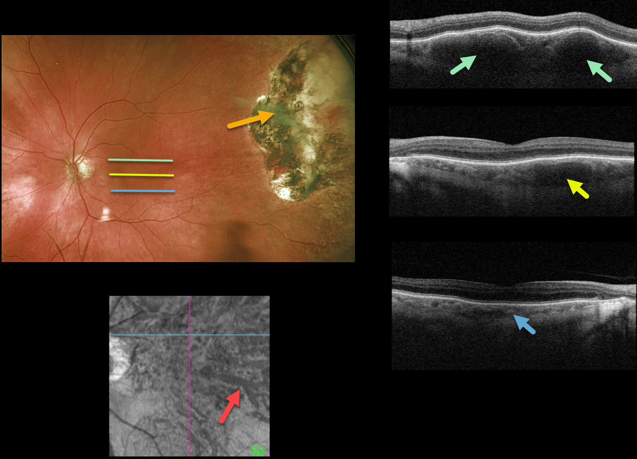
This 85YO male underwent cryotherapy 7 years earlier for a symptomatic hemorrhagic ectopic choroidal neovascularization in his left eye. Optos imaging shows the current appearance of the inactive cryotherapy scarring.
Although the macula has a normal fundoscopic appearance, Triton swept-source OCT scanning is far from normal. B-scans are shown through the top, central, and inferior macula. Although the inferior macular choroid appears relatively normal, a thickened outer choroid (pachyvessels) is noted more centrally, and is huge superiorly. En face imaging through this abnormal choroid is shown.
Learning Points:
The pachychoroid spectrum is a collection of conditions with a thick (“pachy”) choroid. These pachyvessels can cause a range of disorders, including idiopathic central serous chorioretinopathy, pachychoroid pigment epitheliopathy, choroidal excavation, pachychoroid neovasculopathy, polypoidal choroidal vasculopathy, pachydrusen, and peripapillary pachychoroid syndrome.
They are also associated with ectopic neovascularization (aka peripheral exudative hemorrhagic chorioretinopathy), although the choroid in these cases shows increased thickening towards the temporal periphery (see Shroff et al, Retina 2021;41:1518-1525).



