

This 64YO diabetic male presented with asymptomatic retinal findings in his left eye. Vision was 20/40 OU.
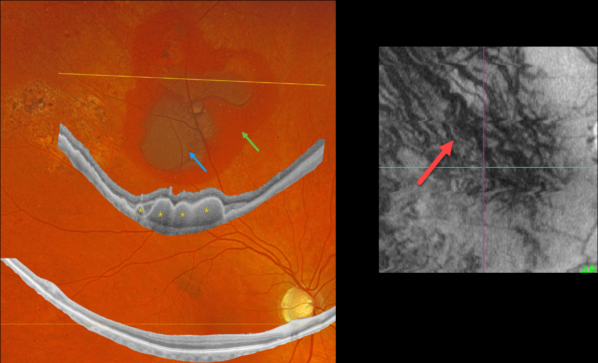
Optos color RGB imaging shows superior subretinal and sub-RPE blood. Optos Silverstone widefield swept-source OCT through this lesion shows prominent multiple choroidal polyps. Subretinal pigmentary clumping temporal to the blood likely represents findings from absorbed subretinal blood. Moderate non-proliferative retinopathy is noted.
En face imaging is quite abnormal with vortex anastomoses and dilated choroidal vessels.
Observation was recommended.
Learning Points:
Pachychoroid, meaning “thick” choroid, is secondary to large, dilated Haller vessels. Increased oncotic/mechanical pressure from these dilated vessels can cause RPE dysfunction, serous detachments, and macular neovascularization.
What defines a ‘pachy’ choroid remains quite unclear (Spaide, Retina 2021;41:231-237), since pachychoroid-related findings can be present despite a ‘normal’ macular choroidal thickness as in our patient.
Spaide recently described venous overload choroidopathy, a unifying concept that better explains the underlying pathogenesis (Progress Retinal Eye Research 2022;86:100973).
The pachychoroid spectrum also includes ectopic neovascularization (aka peripheral exudative hemorrhagic chorioretinopathy), with the choroid showing progressive thickening towards the temporal periphery (Shroff et al, Retina 2021;41:1518-1525).
Our patient’s widefield OCT beautifully shows the choroidal polyps responsible for the peripheral subretinal bleeding.



