

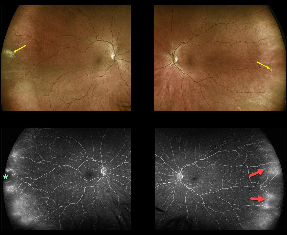
This healthy, asymptomatic 40YO female presented with 20/25 vision bilaterally.
Optos ultra-widefield (UWF) imaging showed far temporal peripheral probable lattice lesions and subtle telangiectasia. The vascular changes, including capillary dropout and telangiectasia, were more evident on fluorescein angiography (FA).
Learning Points:
In 2016, Shah et al described common peripheral retinal vascular findings on Optos UWF FA in otherwise healthy adults (58 eyes, 31 patients) undergoing imaging for unrelated macular pucker or choroidal nevi (Retina 2016;36:1087-1092).
Findings included vessels crossing the horizontal raphe (45%), right-angle vessels (71%), terminal networks (78%), absence of capillary detail (98%), ground glass hyperfluorescence (88%), drusen (34%), and microaneurysms (41%). One or more of these findings were present in most eyes.
Another study by Lu et al. found that normal peripheral retinas typically exhibit granular background fluorescence, with or without a mottled fluorescent band (AJO 2017;173:84-90).



