

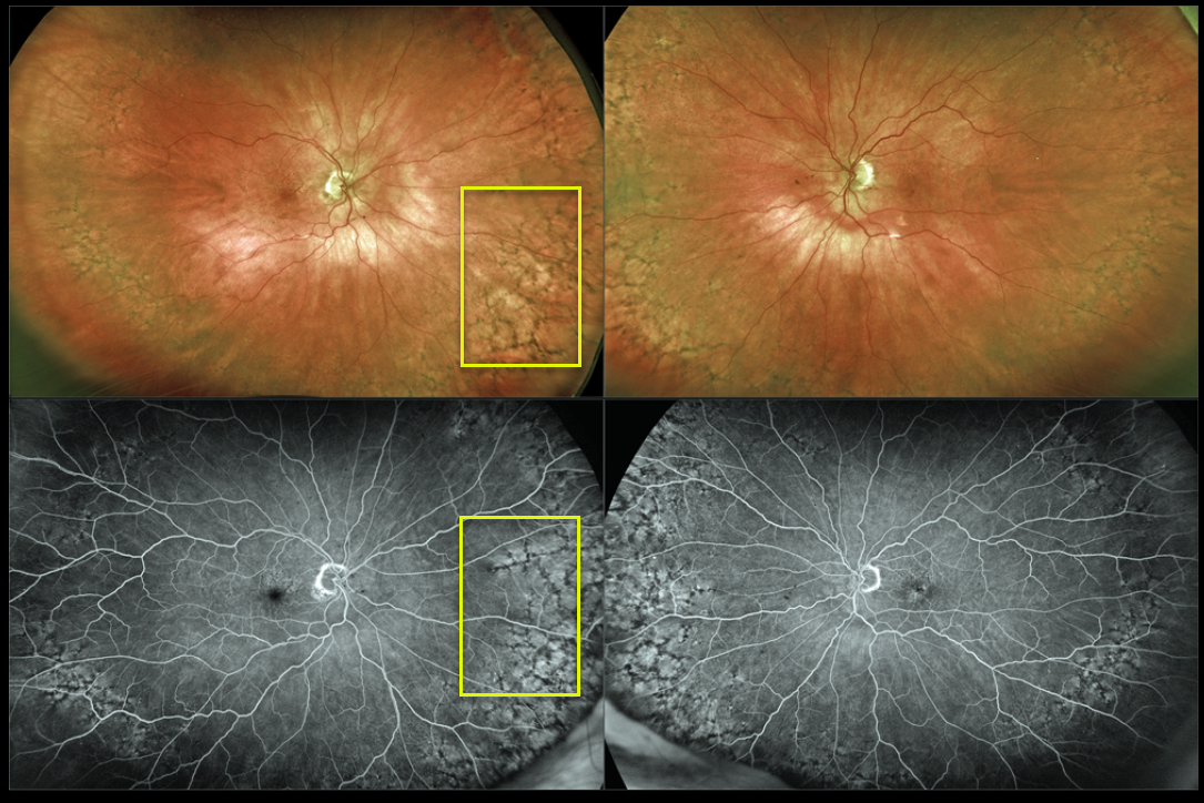
This 87YO female shows prominent bilateral peripheral reticular degeneration of the RPE (PRDRPE) with Optos ultra-widefield imaging and fluoroscein angiography.
The reticular changes at first glance may appear similar to the ‘bone spicules’ found with retinitis pigmentosa, which are caused by disinhibited RPE cells migrating along retinal capillaries following photoreceptor degeneration.
Learning Points:
The findings in PRDRPE are part of the age-related macular degeneration (AMD) spectrum and are subretinal and located in the periphery. A recent meta-analysis by Forshaw et al found PRDRPE to be present in 68-83% of eyes with AMD (Ophthalmology Retina 2019;734-743).
It is associated with decreased dark adaptation, which may help explain why many patients with AMD experience difficulty with night vision (see Lains et al, AJO 2018;190;113-124).



