

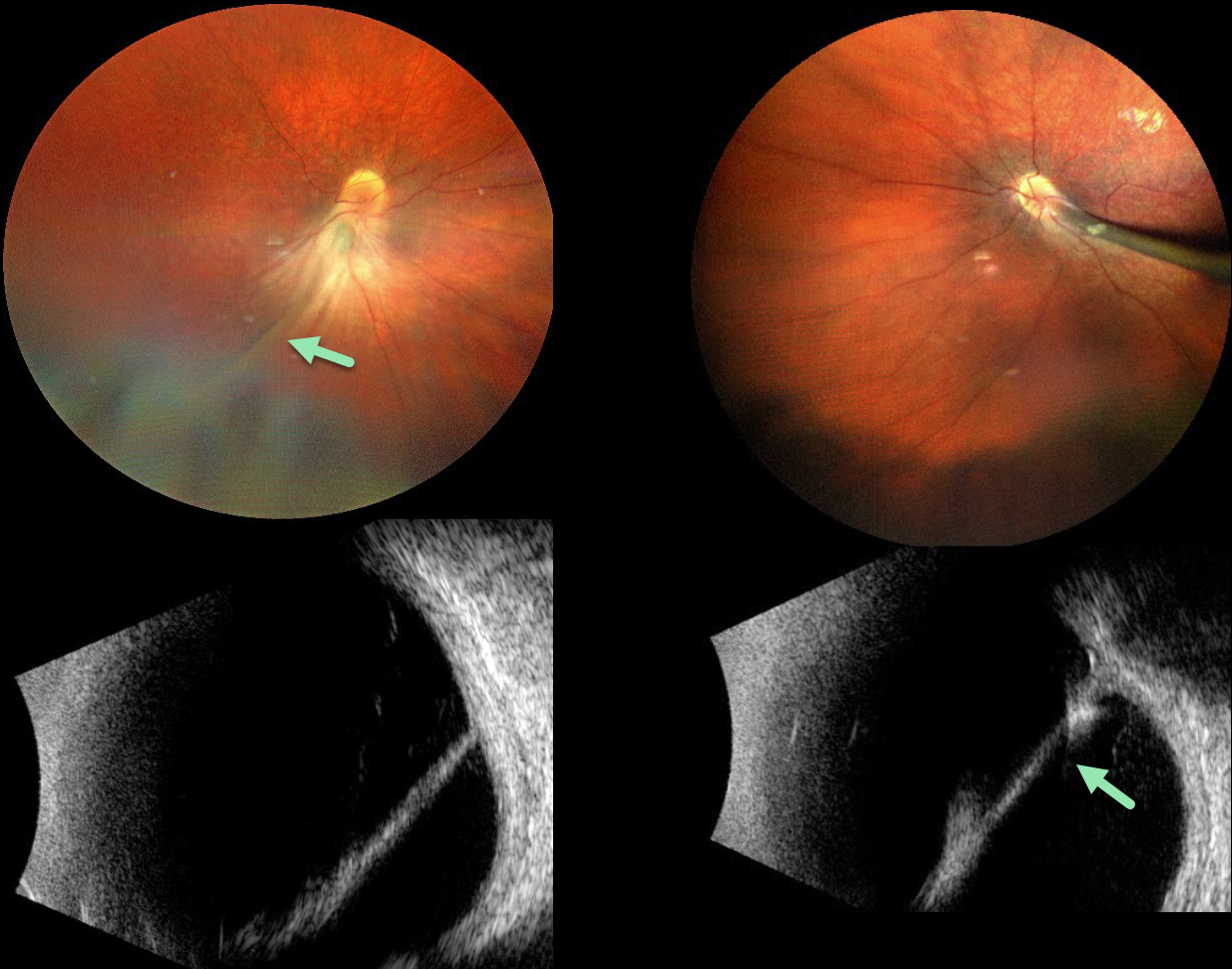
The 9YO girl presented with a known history of persistent fetal vasculature (PFV) since age 4. She was the product of a normal, full-term pregnancy. Vision was 20/400 OU. She also has congenital nystagmus.
Color photography shows bilateral fibrous stalks extending from the optic disc to the posterior lens capsule. The stalks are hyperreflective on B-scan ultrasonography. Falciform folds are also observed. Each retina was otherwise completely attached. She was referred to a pediatric retinal specialist for further management.
Learning Points:
Persistent fetal vasculature (PFV), previously recognized as persistent hyperplastic primary vitreous (PHPV), represents a failure of regression of fetal vessels. It is a common cause of leukocoria and subsequent amblyopia. There is no identifiable genetic association.
Characteristically, the affected eye is microphthalmic with a shallow chamber predisposed to secondary angle closure glaucoma, a varying degree of cataract, and retrolental and/or posterior fibrovascular membranes that can cause retinal traction.
If diagnosed early in life, PFV can be surgically treated with a combined lensectomy and vitrectomy. Anterior-type PFV is associated with better vision than combined or posterior types, and younger age at presentation correlates with improved vision after treatment.
For a review of PFV signs and symptoms, see Goldberg, AJO 1997;124:587-626.



