

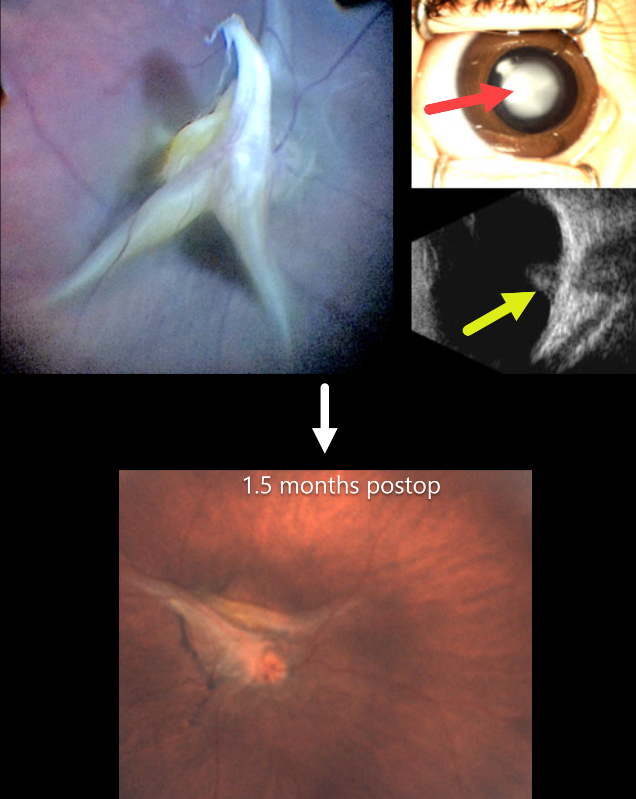
This healthy 4-month-old girl, born full-term, presented with leukocoria in her right eye that her parents recently noticed.
Preoperatively, there was a white central cataract with a clear peripheral zone. B-scan ultrasonography showed a highly reflective stalk that extended from the disc to the periphery with a tent-like elevation over the disc. The left eye was normal.
At surgery, the lens was aspirated with vitrectomy to release all traction. Although the stalk overlying the nerve was trimmed, persistent blood flow was noted as stalk pulsations. The retina remained attached 1.5 months postoperatively.
This case was submitted by the European VitreoRetina Society (EVRS), Gökçen Deniz, and Şengül Özdek .
Learning Points:
Persistent fetal vasculature (PFV), previously recognized as persistent hyperplastic primary vitreous (PHPV), represents a failure of regression of fetal vessels. It is a common cause of leukocoria and subsequent amblyopia. There is no identifiable genetic association.
In PFV, the arterial blood supply from the disc to the anterior segment that is supposed to regress after birth does not. This results in a remnant of the fetal hyaloid artery remaining abnormally patent. The arterial flow from this remnant can cause cataract, intraocular hemorrhages, and retinal detachment, as the abnormal vessel continues to be supplied by systemic arterial pressure.
The main surgical pearl of this case is that, when you have a clear zone around the central opacity of PFV, such eyes usually do not have peripheral retinal extensions through the ciliary body, which allows safe surgical entry through the pars plicata. One still needs to check internally that the incisions are anterior to the peripheral retina, and limbal entry is needed.



