

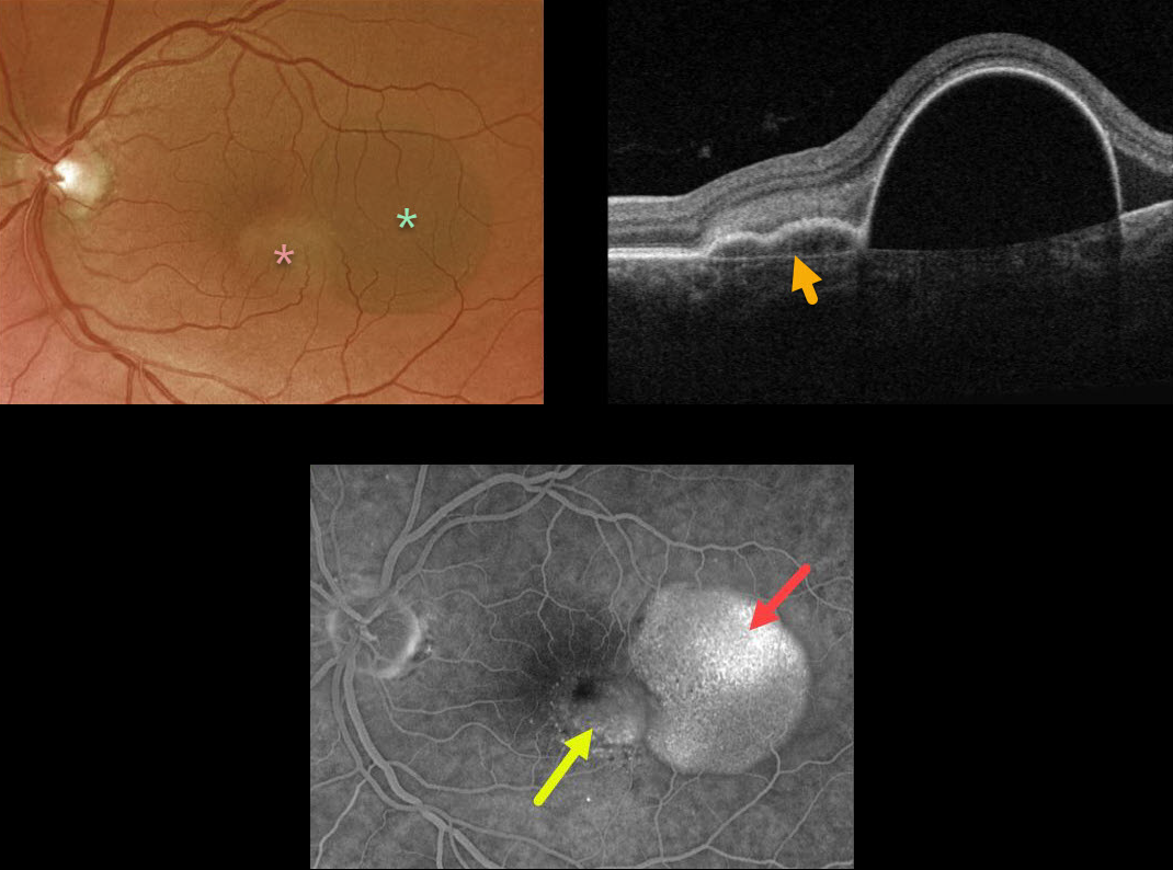
This 57YO female presented with counting-finger vision from a large RPE detachment (PED). The PED was located in the temporal macula, with a smaller notch of irregular RPE elevation more centrally.
Triton swept-source OCT shows a shallow PED at the notch overlying Bruch’s membrane. Optos fluorescein angiography shows late uneven filling of the PED and late speckled staining of the notch.
Anti-VEGF therapy was recommended.
Learning Points:
Originally described by Gass (Retina 1984;4:205-220), a notched PED is a sign of an occult macular neovascularization (MNV). The notch represents a type 1 MNV, which tethers the overlying RPE from the surrounding more highly elevated PED. Hydrostatic leakage from the MNV may contribute to the larger PED.



