

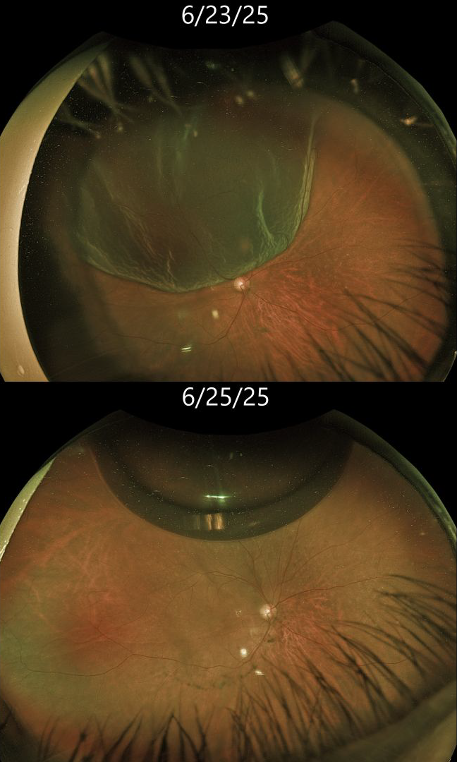
This 76YO female presented with several days of painless vision loss in her right eye. Vision was 20/30.
Optos color RG imaging shows a bullous superotemporal rhegmatogenous retinal detachment (RRD). The macular center was shallowly detached. Scleral depression revealed a single tiny retinal tear anterior to the 11:30 equator. Cryotherapy for this tear, followed by pneumatic retinopexy with 0.2 cc C3F8, was performed. Two days later, vision was 20/30. A single gas bubble is noted superiorly, and the retina is completely attached.
Learning Points:
Pneumatic retinopexy (PnRP) was introduced by Hilton and Grizzard in 1986 as an in-office alternative to traditional retinal reattachment surgery (Ophthalmology 1986;93:626-651). It was initially recommended for detachments with one or more breaks within a single clock hour, located within the superior eight clock hours of the fundus.
Nearly 40 years after its introduction, the role of RRD repair in RRD repair, compared with scleral buckling and vitrectomy, remains controversial. In Canada, where access to operating rooms is more limited than in the US, PnRP is often the initial treatment of choice for appropriate detachments. The Canadian PIVOT trial reported that PnRP offered better final vision, less vertical metamorphopsia, and reduced morbidity compared to vitrectomy (Ophthalmology 2019;126:531-539). However, in the US, PnRP is not as popular except on a late Friday afternoon (we’re only partially kidding). A recent critical review (Chronopoulos et al, Surv Ophthalmology 2021;66:585-593) argued against PnRP for initial treatment due to the need for “multiple procedures that largely negates its potential cost savings and subjects the patient to prolonged stress and disability…” A recent cost-utility analysis surprisingly found PPV to be the most cost-effective primary procedure (Teja et al, AJO 2023;255:141-154).
We personally recommend PnRP for patients with mobile superior RRD with superior breaks. Patients must be able to properly position their head. We must be able to visualize the retinal periphery well to ensure there are no inferior breaks, and ideally, we prefer that the macula be detached in case a failed PnRP converts a macula-on RRD to a macula-off RRD.
We therefore felt that our patient was a perfect candidate for PnRP. The PIVOT trial also supported our decision, with a recent post hoc analysis showing similar single-surgery success with PnRP (88%) vs PPV (91%) for single-break primary RRD (Pecaku et al., Br J Ophthalmol 2025;109:113-118).



