

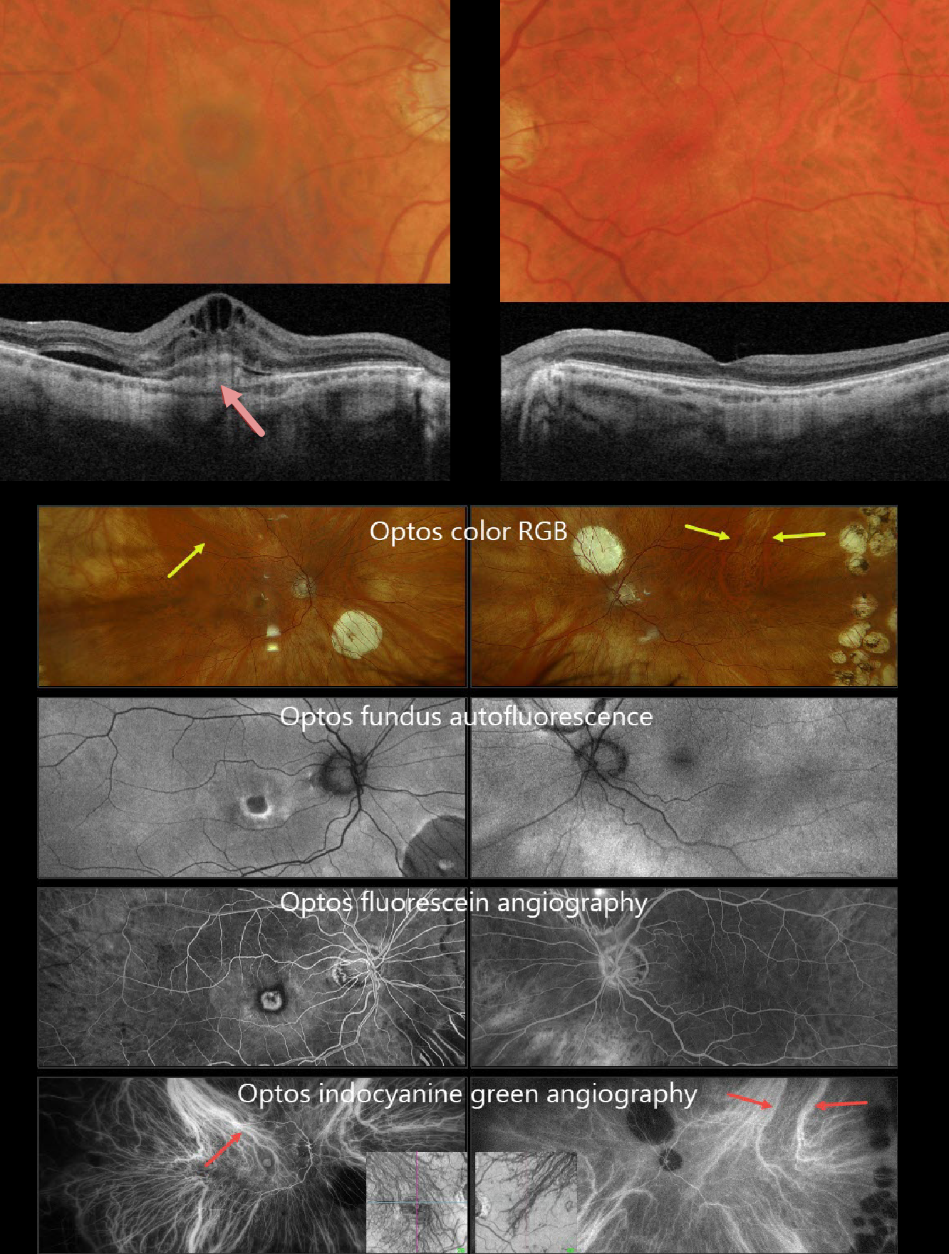
This 71YO white male underwent successful bilateral vitrectomies for rhegmatogenous retinal detachments by us years earlier. He had since moved out of state and was recently diagnosed with neovascular AMD in his right eye, which was treated with an initial anti-VEGF injection one month earlier. After moving back to our area, vision was 20/400 OD and 20/20 in his normal OS.
Triton color photography shows a poor foveal reflex with a surrounding subretinal pigment ring. Barely visible, smaller drusen are noted more peripherally. Swept-source OCT shows a probable type 1 (below the RPE) macular neovascularization (MNV) with overlying subretinal hyperreflective material, intraretinal and subretinal fluid. There are multiple smaller drusen in his left macula. Although the choroid appears thinned bilaterally, the vessels that are present mostly span the entire choroidal thickness.
Optos fundus autofluorescence (FAF) shows the MNV to be hypo-FAF with a rim of surrounding hyper-FAF (image 2). The MNV is hyperfluorescent on fluorescein angiography, surrounded by a rim of blockage from the subretinal pigment. The choroid is markedly abnormal on indocyanine green angiography, revealing that each macula is drained by the superotemporal choroidal vortex. These dilated choroidal vessels are also visible on color RGB imaging.
Learning Points:
Polypoidal choroidal vasculopathy (PCV) is a variant of type 1 macular neovascularization. PCV is likely distinct from age-related macular degeneration, occurring in a different population (more common in African Americans and Asians) and characterized by a relative lack of drusen and a thicker choroid. PCV presents differently in Caucasians, with multiple phenotypes that can include coexisting drusen, the presence or absence of a branching choroidal neovascular network, or background central serous chorioretinopathy findings (Sirks et al., Ophthalmology Retina 2025;9:994-1004).
PCV is part of the pachychoroid spectrum, more recently termed venous overload choroidopathy, which appears to be caused by relative vortex outflow resistance (Spaide et al., Progress Retinal Eye Research 2022;86;100973). The choroidal vortex system usually respects the horizontal meridian; anastomoses that cross it, as occurred in our patient, are characteristic of these eyes.
Historically, these lesions were believed to be somewhat resistant to anti-VEGF monotherapy and often required combined treatment with photodynamic therapy. More recent data from the PLANET study suggest that Eylea monotherapy is also effective (AJO 2019;204:80-89).
Given the lack of response to initial anti-VEGF therapy, intravitreal Eylea was given, followed by PDT 1 week later. Six weeks following PDT, vision remained stable, but the OCT showed marked improvement. We will continue to follow him closely.



