

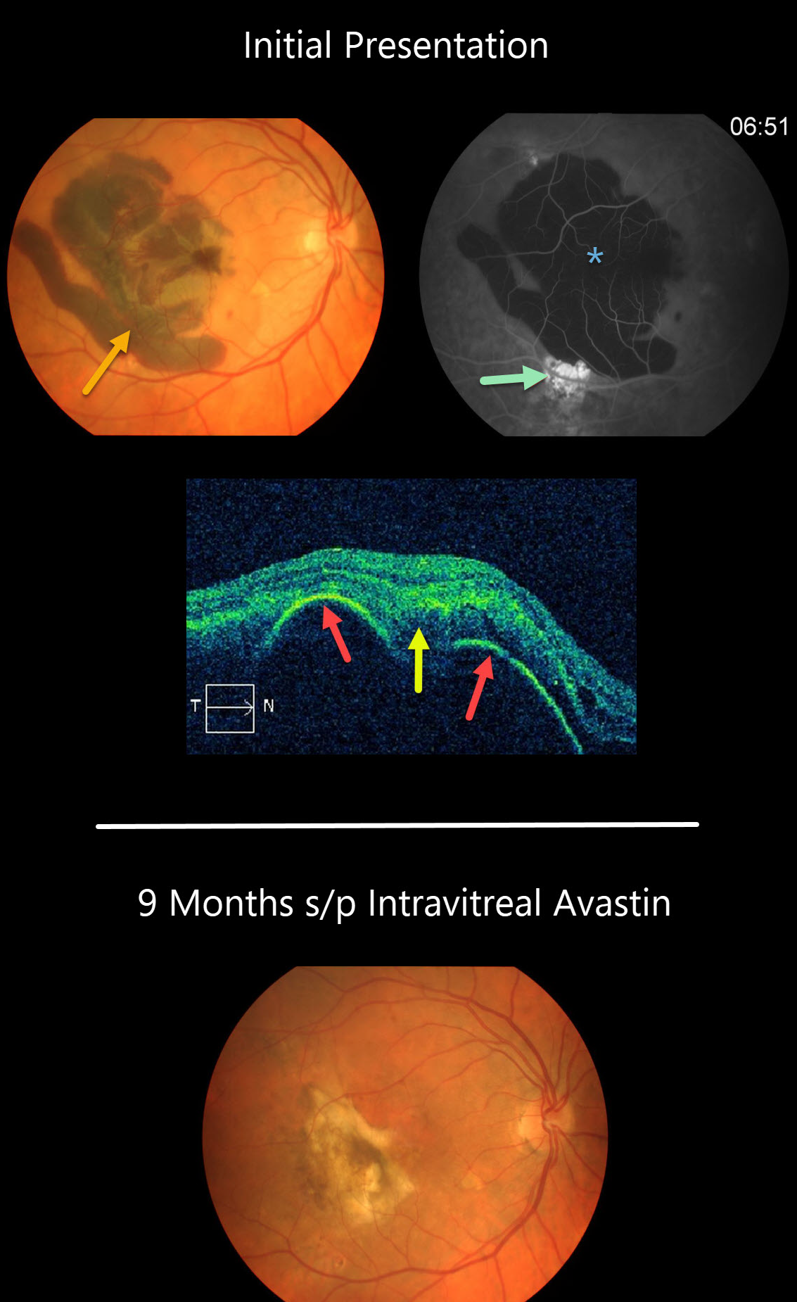
This patient presented with a large acute submacular hemorrhage in the right eye.
The blood caused diffuse blockage on fluorescein angiography, and the source of the blood may have been from the area of subretinal hyperfluorescence inferiorly.
OCT scanning shows a large retinal pigment epithelium (RPE) detachment with notched thumb-like projections with subretinal hyperreflectivity from the blood.
Our patient’s subretinal blood resolved following several intravitreal Avastin injections.
Learning Points:
Polypoidal choroidal vasculopathy (PCV) is a variant of type 1 macular neovascularization. PCV is likely distinct from age-related macular degeneration, occurring in a different population (more common in African Americans and Asians) and characterized by a relative lack of drusen and a thicker choroid.
The sub-RPE vessels were classically characterized using indocyanine green angiography, although recent literature suggests that OCT is just as effective at diagnosing these vascular polyps. Historically, these lesions were believed to be somewhat resistant to anti-VEGF monotherapy and often required combined treatment with photodynamic therapy. More recent data from the PLANET study suggest that Eylea monotherapy is also effective (AJO 2019;204;80-89).



